Mandibular Third Molar CBCT: Reading the IAN Canal

A mandibular third molar CBCT is indicated when the panoramic radiograph shows signs of proximity to the inferior alveolar canal, surgery has already been decided, and the result could change the technique or the decision [1]. Its value is not a lower rate of inferior alveolar nerve (IAN) injury per se, which randomized trials have not shown [2],[3],[4], but showing whether bone separates root and canal, on which side the canal runs and whether its cortical border is intact. That is what separates conventional removal from coronectomy.
Key points
- The European SEDENTEXCT guidelines (Radiation Protection No. 172) state that CBCT may be indicated (grade C) when conventional radiographs suggest a direct relationship between the third molar and the canal and removal has been decided; routine use is not recommended [1].
- The panoramic signs associated with nerve injury are darkening of the root, interruption of the canal's cortical outline and diversion of the canal [5],[1].
- In a multicentre trial of 268 patients and 320 high-risk third molars, temporary IAN injury was 6.3% and did not differ between CBCT and panoramic imaging (p = 0.64) [2].
- In the same trial, a lingual canal position and canal narrowing at the root contact area were significant risk factors [2].
- In a series of 145 third molars, all 7 nerve injuries (4.8%) occurred where the canal lacked corticalization [6].
- No bony separation between tooth and canal on CBCT was the main factor behind choosing coronectomy (OR 56.8) in an audit of 1,437 third molars [7].
When should you order a mandibular third molar CBCT?
The panoramic radiograph remains the first-line exam. Rood and Shehab's classic study assessed seven radiographic signs and found three significantly related to nerve injury [5]; SEDENTEXCT summarizes them as darkening of the root, interruption of the canal wall and diversion of the canal [1]. When none is present, CBCT rarely adds anything that would change surgery.
When proximity signs are present, the right question is not "is there contact?" but "will CBCT change what I am going to do?". The guideline is explicit: if coronectomy has already been planned on conventional radiographs, CBCT is unnecessary, and routine use is not justified because post-surgical dysaesthesia is rare in experienced hands [1]. In practice, the strongest indication is a case with proximity signs in which the surgeon is weighing full removal, coronectomy or a modified technique (sectioning, direction of elevation).
Acquisition protocol
A small FOV centred on the third molar region answers this question, using a voxel size that resolves the canal's cortical border without unnecessary dose. Dose and FOV reasoning is covered in our article on CBCT radiation dose.
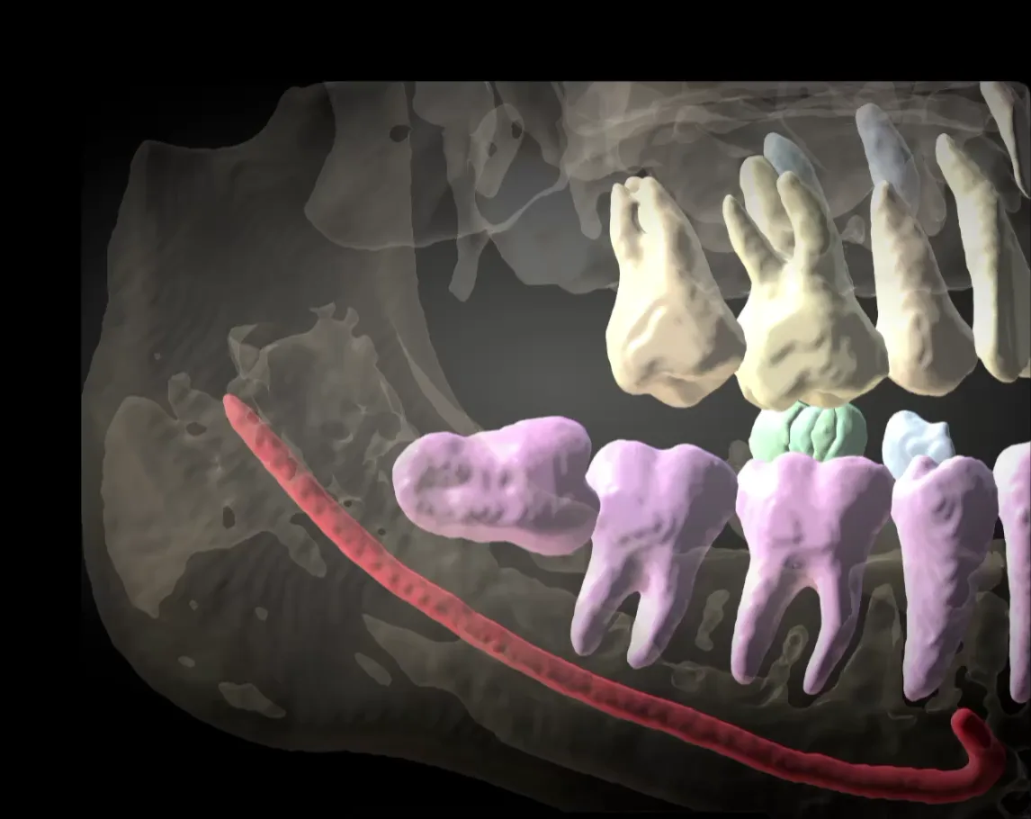
What to assess: mandibular third molar and inferior alveolar canal
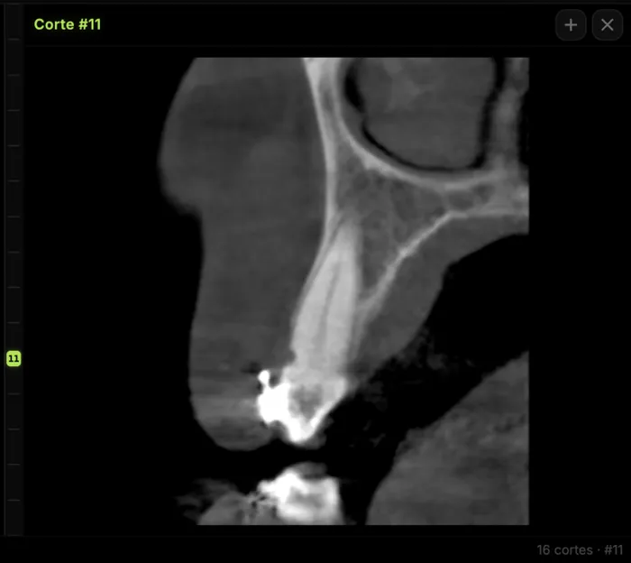
Read on cross-sectional slices perpendicular to the arch curve, scrolling through the tooth from mesial to distal, and confirm on axial slices and the panoramic reconstruction. A single slice is never enough: contact may be visible on one slice and disappear on the next, so the series must cover the whole apical third.
| Parameter | How to read it on CBCT | Why it matters | What to report |
|---|---|---|---|
| Intervening bone | Look for trabecular bone or cortex between root and canal on every apical slice | Its absence weighed most in the coronectomy decision [7] | "Direct contact without intervening bone" or "bony separation of X mm" |
| Canal position | Buccal, lingual, inferior (apical) or interradicular relative to the roots | A lingual position was a risk factor for temporary injury [2] | Side and root involved (mesial/distal) |
| Corticalization | Continuity of the hyperdense line outlining the canal at the contact area | All injuries in Ueda's series lacked cortication [6] | Cortex intact, thinned or absent |
| Shape and calibre | Round/oval, teardrop or dumbbell canal; narrowing at the contact area | Shape was associated with injury in non-corticated cases [6]; narrowing was a risk factor [2] | Canal shape and any narrowing or root grooving |
| Roots | Number, curvature, dilaceration, fusion, canal passing between roots | Drives sectioning and direction of elevation | Root morphology and relationship with the canal |
| Surroundings | Lingual cortex, second molar (distal resorption, caries), follicle, associated lesions | Risk of lingual displacement and second molar damage | Findings from the whole volume, not just the tooth |
A 6-step reading protocol
- Draw the panoramic curve through the centre of the ridge and check that the canal can be followed continuously from the mandibular foramen.
- Generate thin, contiguous cross-sections covering the entire third molar, from mesial of the mesial root to distal of the distal root.
- Identify the canal before it reaches the tooth (where nothing overlaps) and follow it into the contact area.
- On each apical-third slice: intervening bone, canal side, cortex and shape.
- Confirm on axial slices and on an oblique slice aligned with the root axis when the root is inclined.
- Review the rest of the volume: second molar, lingual cortex, follicle and any incidental finding.
For canal anatomy and variants (bifid and retromolar canals, anterior loop), see our guide to the mandibular canal and inferior alveolar nerve on CBCT.
Common pitfalls. Judging contact on the CBCT panoramic reconstruction, which sums several millimetres of thickness; reading a single cross-section; using a window so narrow that the canal cortex "disappears"; ignoring beam hardening from metallic restorations on the second molar; and accepting an automatic canal tracing or AI segmentation without checking it.
Does CBCT reduce inferior alveolar nerve injury?
Randomized evidence is consistent: adding CBCT to the panoramic radiograph does not routinely reduce IAN injury.
| Study | Design | Sample | Main result |
|---|---|---|---|
| Ghaeminia et al., 2015 [2] | Multicentre RCT | 268 patients, 320 high-risk molars | Temporary injury 6.3%; no difference CBCT vs panoramic (p = 0.64) |
| Del Lhano et al., 2020 [3] | Systematic review and meta-analysis | 4 RCTs | RR 1.23 (95% CI 0.75–2.02); moderate certainty; no significant difference |
| Robbins et al., 2022 [4] | Systematic review | 7 RCTs | Only 1 of 7 trials showed fewer injuries with CBCT; temporary injury 2.3–14.8%; permanent 1.1% (10/888) |
| Matzen et al., 2020 [7] | 3-year retrospective audit | 1,437 molars, 917 patients | 29% referred for CBCT; no intervening bone → coronectomy (OR 56.8) |
Robbins et al. recommend reserving CBCT for high-risk cases where 3D imaging may change the decision or technique [4].
From finding to decision: removal, coronectomy or monitoring
CBCT earns its place when it changes the plan. In the Danish audit by Matzen et al., of 840 third molars operated on, 152 were managed with coronectomy and 688 with full removal; no bony separation between tooth and canal was the finding most associated with choosing coronectomy [7]. Coronectomy is backed by a randomized trial: in 349 third molars, inferior dental nerve deficit occurred in 9 patients in the removal group versus 1 in the coronectomy group (p = 0.023), with 16 failed coronectomies ending in root removal [8].
- Intervening bone on every slice: conventional removal; CBCT provides reassurance rather than a change of technique.
- Contact with intact cortex, buccal or inferior canal: removal with sectioning and elevation planned away from the canal.
- Contact without bone, absent cortex, lingual, interradicular or narrowed canal: discuss coronectomy or a modified technique with the surgeon and document the risk in the consent.
The final decision is clinical and weighs age, associated pathology and the team's experience. For the related surgical planning workflow, see CBCT for third molar extraction planning.
What the report should include and how to communicate it
The report should answer the clinical question in the first line of the impression and then describe the rest of the volume:
- Technique: unit, FOV, voxel size.
- Tooth: position, angulation, number and morphology of roots.
- Relationship with the canal: contact yes/no, intervening bone, canal side, cortex, shape and narrowing, with the reference slice number.
- Surroundings: second molar, lingual cortex, follicle, associated lesions.
- Incidental findings in the rest of the volume.
Sample wording: "Distal root of tooth 48 in direct contact with the mandibular canal, without intervening bone on slices 41–44; canal in a lingual position, with no cortical outline at the contact area and narrowing of its calibre." The choice of technique rests with the surgeon.
How to do it in CBCTHub
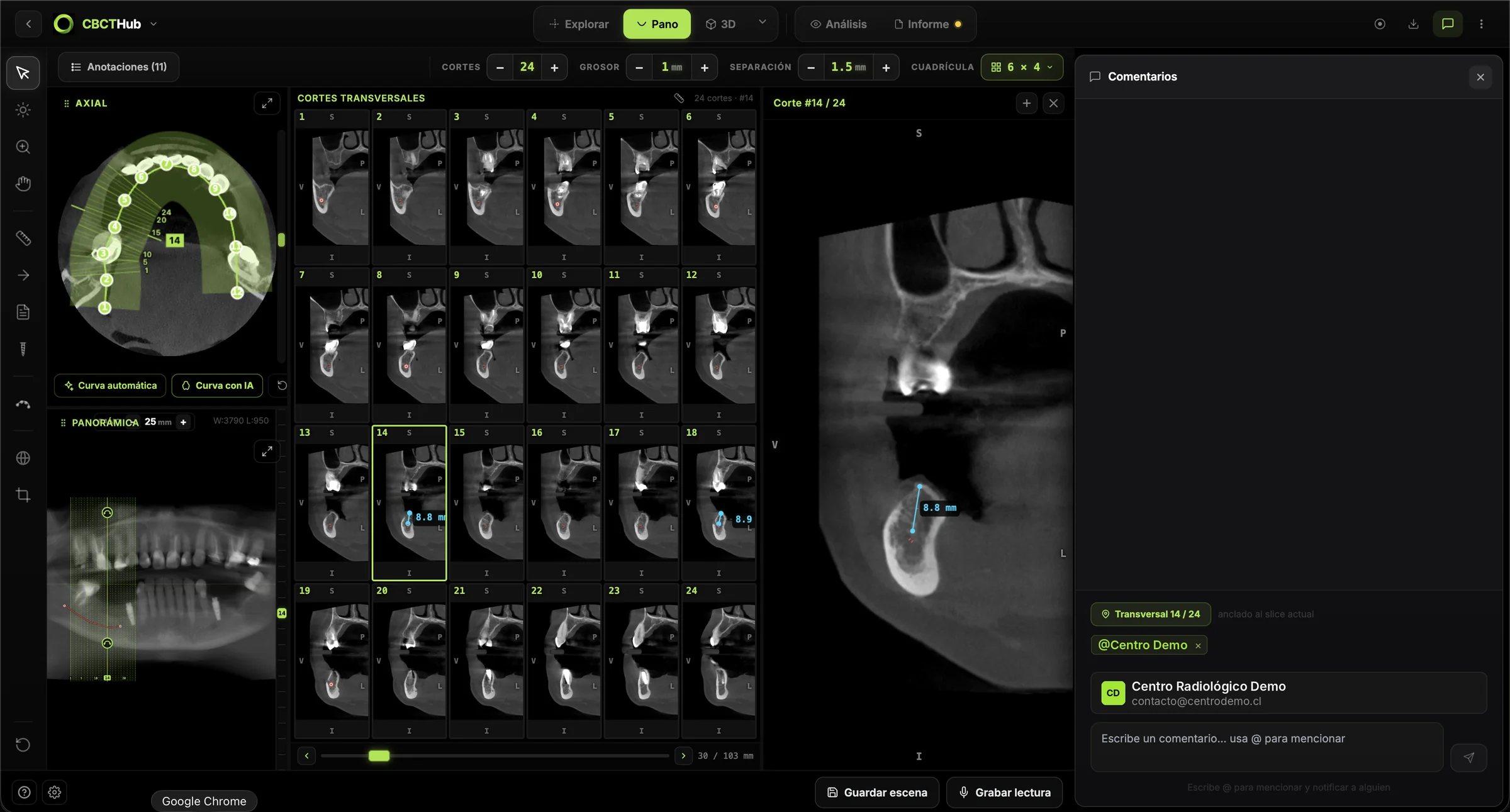
CBCTHub lets you read, report and share a third molar CBCT in the browser, with no install and no CDs; DICOM is processed locally and Carestream, Planmeca, Vatech, Sirona, Morita, NewTom and other units are supported.
- Open the study and generate the panoramic with the automatic curve; adjust it manually if the ridge is asymmetric.
- Scroll through the cross-sections of the third molar and use oblique slices when the root is inclined.
- Measure root–canal separation or lingual cortex thickness in mm.
- Use AI segmentation and the 3D view to show the root–canal relationship, always verifying it on the slices.
- Pin comments to key slices, write the report in the editor and build PDF image plates.
- Share the study by link (with a PIN if needed) or present it live to the surgeon.
Radiologists and imaging centres: deliver the study and report to referring dentists by link, with no CDs, on a study page branded with your centre. Surgeons and dentists: open and measure the CBCT your imaging centre sends you, right in the browser. Try the viewer free, with no account, at cbcthub.com/viewer, or create an account at cbcthub.com.
Conclusion
A mandibular third molar CBCT does not protect the nerve by itself; the decision made with it does. Order it when the panoramic shows proximity signs and the result could change the technique, read it on contiguous cross-sections, and explicitly report intervening bone, canal position, cortex and shape. This content is intended for professionals and does not replace the clinical judgement of the treating team.
Frequently asked questions
When is CBCT indicated before removing a mandibular third molar?
When the panoramic radiograph shows signs of proximity to the mandibular canal, such as darkening of the root, interruption of the canal's cortical outline or diversion of the canal, and surgery has already been decided. SEDENTEXCT rates this as a possible grade C indication and advises against routine use. If coronectomy is already planned on conventional radiographs, CBCT will not change management.
Does CBCT reduce the risk of inferior alveolar nerve injury?
Not routinely. A multicentre trial of 320 high-risk third molars found no difference in temporary nerve injury between CBCT and panoramic imaging, and two systematic reviews of randomized trials confirm this with moderate certainty. The value of CBCT lies in identifying the high-risk case and changing the surgical strategy, for instance towards coronectomy.
Which CBCT finding weighs most when choosing coronectomy?
The absence of bone between the tooth and the mandibular canal. In an audit of 1,437 third molars, this finding was the main factor associated with the decision to perform coronectomy, with an odds ratio of 56.8. A lingual canal position, loss of the canal cortex and narrowing at the contact area strengthen the indication and should be reported explicitly.
What does a non-corticated mandibular canal mean?
It means the hyperdense line that outlines the canal is not visible where it meets the root, so the neurovascular bundle lies directly against the tooth. In a series of 145 third molars, every nerve injury occurred in cases without canal cortication. Before reporting it, confirm the finding on several slices and with an appropriate window setting.
Which FOV is appropriate for assessing a mandibular third molar?
A small FOV centred on the third molar region is enough to answer the surgical question and keeps the dose down. A bilateral field is justified only when both third molars are scheduled for surgery and show proximity signs. Whatever the FOV, the radiologist is responsible for describing the entire acquired volume, including incidental findings.
References
- European Commission. Radiation Protection N° 172: Cone beam CT for dental and maxillofacial radiology. Evidence-based guidelines (SEDENTEXCT). Luxembourg: Directorate-General for Energy; 2012. energy.ec.europa.eu
- Ghaeminia H, Gerlach NL, Hoppenreijs TJ, Kicken M, Dings JP, Borstlap WA, et al. Clinical relevance of cone beam computed tomography in mandibular third molar removal: a multicentre, randomised, controlled trial. J Craniomaxillofac Surg. 2015;43(10):2158-67. doi:10.1016/j.jcms.2015.10.009
- Del Lhano NC, Ribeiro RA, Martins CC, Assis NMSP, Devito KL. Panoramic versus CBCT used to reduce inferior alveolar nerve paresthesia after third molar extractions: a systematic review and meta-analysis. Dentomaxillofac Radiol. 2020;49(4):20190265. doi:10.1259/dmfr.20190265
- Robbins J, Smalley KR, Ray P, Ali K. Does the addition of cone-beam CT to panoral imaging reduce inferior dental nerve injuries resulting from third molar surgery? A systematic review. BMC Oral Health. 2022;22(1):466. doi:10.1186/s12903-022-02490-x
- Rood JP, Shehab BA. The radiological prediction of inferior alveolar nerve injury during third molar surgery. Br J Oral Maxillofac Surg. 1990;28(1):20-5. doi:10.1016/0266-4356(90)90005-6
- Ueda M, Nakamori K, Shiratori K, Igarashi T, Sasaki T, Anbo N, et al. Clinical significance of computed tomographic assessment and anatomic features of the inferior alveolar canal as risk factors for injury of the inferior alveolar nerve at third molar surgery. J Oral Maxillofac Surg. 2012;70(3):514-20. doi:10.1016/j.joms.2011.08.021
- Matzen LH, Villefrance JS, Nørholt SE, Bak J, Wenzel A. Cone beam CT and treatment decision of mandibular third molars: removal vs. coronectomy—a 3-year audit. Dentomaxillofac Radiol. 2020;49(3):20190250. doi:10.1259/dmfr.20190250
- Leung YY, Cheung LK. Safety of coronectomy versus excision of wisdom teeth: a randomized controlled trial. Oral Surg Oral Med Oral Pathol Oral Radiol Endod. 2009;108(6):821-7. doi:10.1016/j.tripleo.2009.07.004
Try CBCTHub for free
Upload, view, and share DICOM scans in the cloud. Nothing to install.
Create free accountRelated articles

CBCT indications in dentistry: when to order and when not
CBCT indications in dentistry per SEDENTEXCT, EADMFR and ADA: when to order a scan, when not to, which FOV to choose and what a referral must include.

Endodontic CBCT indications: a guide to complex cases
Endodontic CBCT indications per AAE/AAOMR: which complex cases justify a scan, which FOV to use, what to report and where CBCT falls short. For clinicians.

CBCT transverse analysis: an orthodontic guide
CBCT transverse analysis: how to measure basal width and molar inclination to tell skeletal deficiency apart from dentoalveolar compensation.