Online CBCT viewer
View CBCT scans with nothing to install
CBCTHub is a CBCT viewer that runs in the browser. Open the DICOM folder your imaging center delivered, scroll through axial, coronal and sagittal slices, draw the panoramic curve, measure in millimeters and show it to your patient on the same screen. No software to download, no licenses, and the scan never leaves your computer.
No account or card · Works on Mac, Windows, iPad and phone
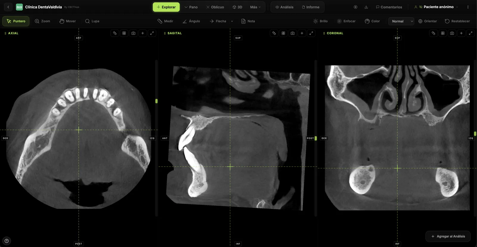
How it works
Drag the folder in. That's it.
No installation, import or conversion. The scan opens exactly as it was delivered to you.
Drop the DICOM
Drag the DICOM folder or ZIP from the imaging center onto the browser window. From a USB drive, a CD or the WeTransfer download.
Full skull, maxilla, mandible or TMJ
Opens in seconds
The browser rebuilds the volume on your own computer. MPR slices, panoramic and 3D are ready without waiting for an upload.
Nothing is sent to any server
Measure and show
Distances, angles, mandibular nerve, cross-sections. Turn the screen and your patient understands what you are explaining.
The same tools as desktop software
Tools
Everything you expect from a workstation, in the browser
The same tools you already use, with nothing to install and from any computer.
- Panoramic Curve

Panoramic curve
Trace the arch, get a panoramic in real time. Adjust thickness from 1 to 40 mm and regenerate on the fly.
- Standard Slices
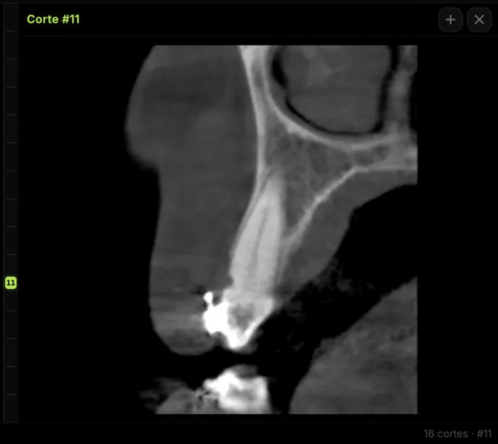
Cross-sections
Perpendicular slices along the panoramic curve, generated automatically. Bone width and nerve proximity at a glance.
- Oblique Slice
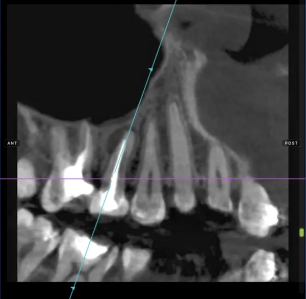
Oblique slices
Slices at any angle with free rotation on three axes. For root canals and fractures.
- Measure
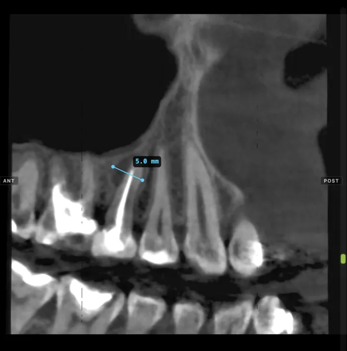
Measurements
Distances with voxel-level millimetric precision. For implants and lesion assessment.
- Implant
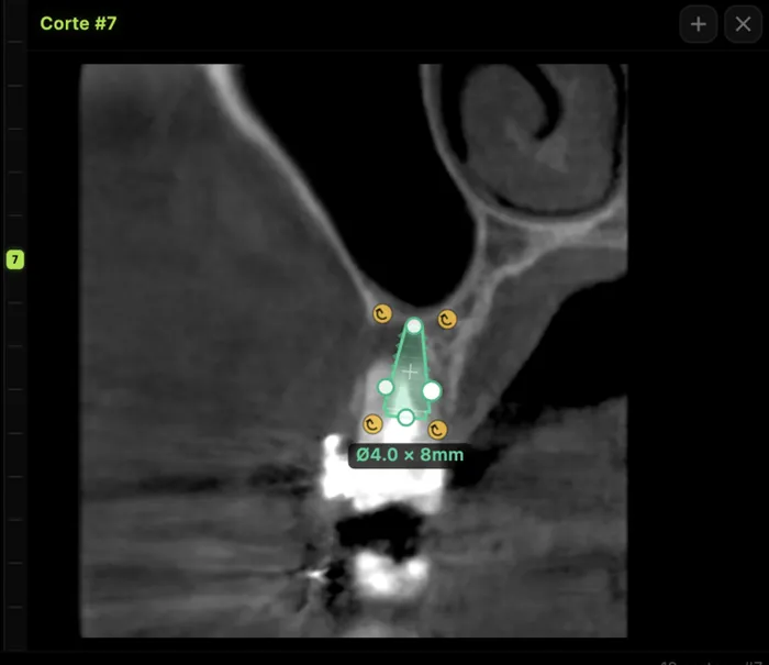
Implant planning
Bone availability, distance to the nerve and bucco-lingual width to plan the ideal implant position.
- Nerve
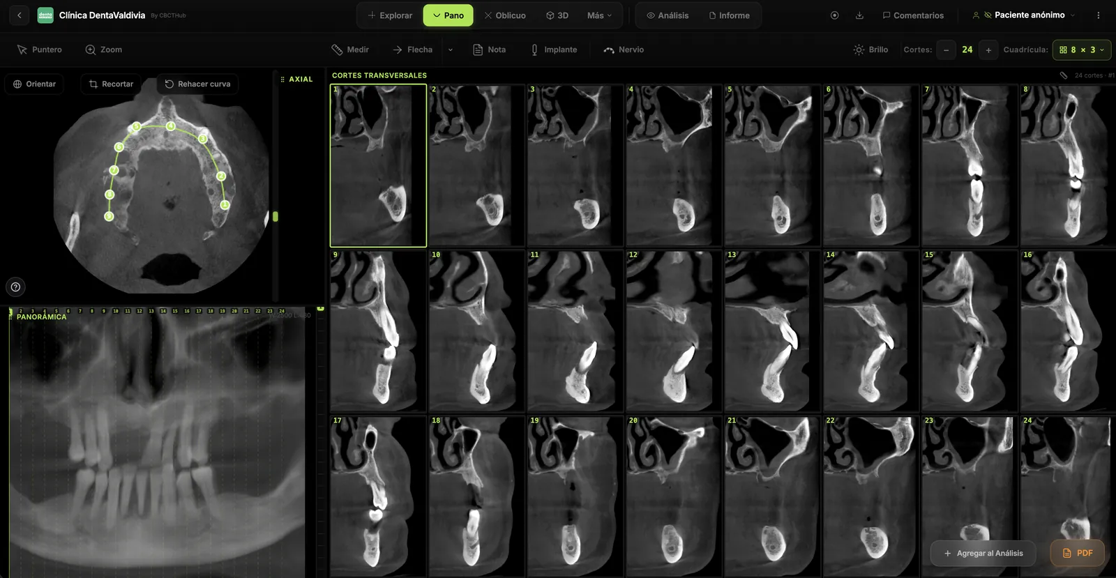
Inferior alveolar nerve tracing
Mark the mandibular canal and see it on every cross-section and on the panoramic view.
- 3D
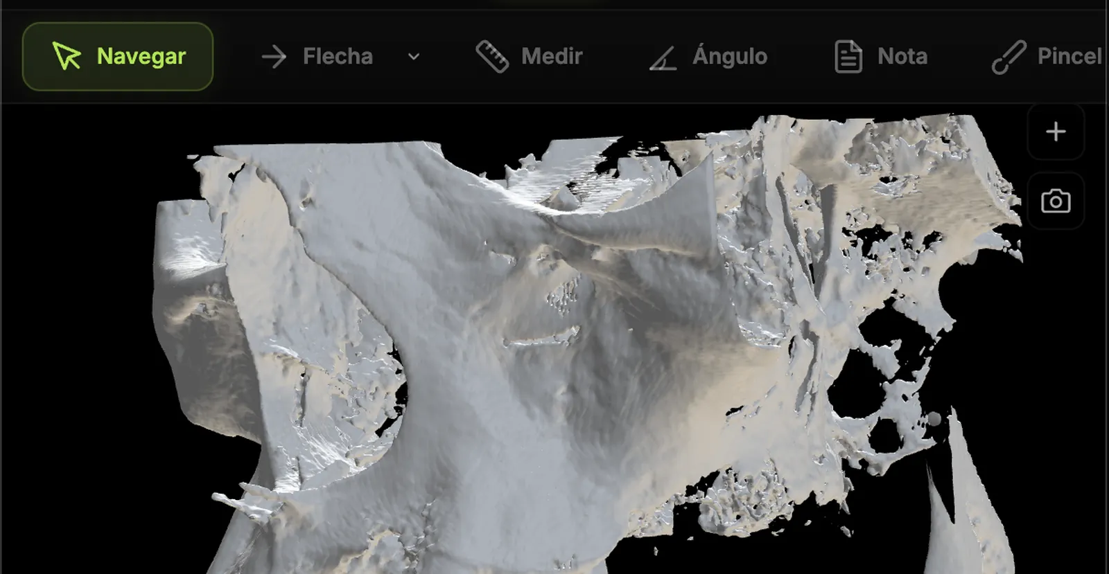
3D reconstruction
Volumetric render of the CBCT with free rotation and volume cropping.
- Angle
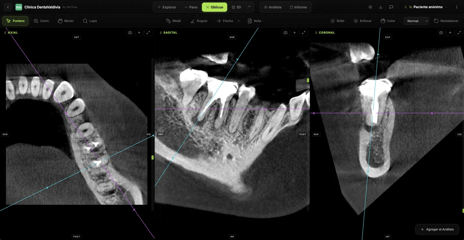
Angles, arrows and notes
Measure angles, point out findings with arrows and leave slice-level notes that travel with the scan.
Privacy
The scan never leaves your computer.
The free viewer processes the DICOM in your browser, with your own CPU and graphics card. There is no upload, no server in between and no sign-up: close the tab and nothing is left anywhere.
Use cases
Every specialty, one viewer
From implant planning to fracture workup — one tool handles it all.
Implantology
Available bone, distance to the alveolar nerve and implant position.
Cross-sectionsNerveImplantOrthodontics
Root relationships, airway and facial symmetry.
MPRPanoramic3DEndodontics
Extra canals, root fractures and periapical lesions.
Oblique sliceZoomMeasureOral & maxillofacial surgery
Fractures, bone pathology and impacted third molars.
MPR3DAngleGuide coming soonTMJ disorders
Both condyles side by side, perpendicular to the condylar axis.
TMJ moduleCompareMeasureGuide coming soon
Each case opens a guide with screenshots, protocol and what to look for in the scan.
What's free and what isn't
The viewer is free and comes with every plan.
What you pay per month is cloud storage. And if you would rather not use the cloud, there is Desktop Viewer: the viewer installed on your computer, with a library and saving, for a one-time payment.
The viewer: what you open your scans with
In the browser
Web viewer
$0 forever
- ✓Every tool: MPR, panoramic, cross-sections, oblique, measurements, nerve, 3D
- ✓Local processing: nothing is uploaded unless you decide
- ✓Included in every plan, the free account too
- ✓Mac, Windows, iPad and phone
Installed on your computer
Desktop Viewer
One-time payment coming soon · no monthly fee
- ✓Everything in the web viewer, offline
- ✓Local library: search by patient and reopen in one click
- ✓Your work is saved: measurements, nerve, curve, notes and scenes stay with the scan
- ✓Export screenshots, slices and a PDF of slices with your logo
- ✓Double-click the DICOM folder or ZIP and it opens
The cloud: where your scans live
To keep your scans in the cloud
Pro
$39 per month · cancel anytime
- ✓Desktop Viewer included while the plan is active
- ✓350 GB of storage for your scans and X-rays
- ✓Link to share with the patient or the specialist
- ✓PDF reports with your logo and brand
- ✓Live presentation to explain a scan remotely
- ✓Priority support
The rule is simple
Desktop Viewer is included in every paid plan (Pro, Clinic and Ultra).
If you want neither a subscription nor the cloud, buy Desktop Viewer once and it is yours. Works with the free account or no account at all.
Prices in US dollars; the amount depends on your region. See all plans → · Running an imaging center? This is your page →
Frequently asked questions
What people ask before opening their first scan.
Do I need to install anything or create an account?+
No. The CBCT viewer opens in the browser (Chrome, Safari, Edge or Firefox) and asks for no sign-up to view, measure or explore a scan. You only need an account if you want to keep scans in the cloud or share them by link.
Which formats does it open?+
DICOM folders (.dcm) exactly as any CBCT unit exports them: Carestream, Planmeca, Vatech, Sirona, Morita, NewTom, i-CAT, KaVo, Acteon and others. It also opens a ZIP with the DICOM inside and STL models to overlay on the volume.
Is the scan uploaded to the internet?+
In the free viewer, no: the DICOM is processed on your own computer and never leaves it. It is only uploaded if you decide to save it to your account, and then it travels and is stored encrypted.
Which tools does it have?+
Synchronized axial, coronal and sagittal slices, panoramic curve with cross-sections, oblique reconstruction, millimeter and angle measurements, mandibular nerve tracing, annotations and arrows, 3D reconstruction, side-by-side comparison of two scans and STL overlay. The same you would expect from desktop software.
Can I use it to plan implants?+
Yes. The panoramic curve generates cross-sections perpendicular to the ridge, where you measure bone height and width and the distance to the nerve you traced. It is the usual workflow before guided surgery.
Does it work on iPad or phone?+
Yes. The viewer runs in any modern browser, including iPad and phones. For working through a full scan we recommend a large screen, but for showing a slice to a patient the iPad works very well.
Is there a one-time-payment version, without a subscription?+
Yes, Desktop Viewer: the viewer installed on your computer (Mac and Windows), with a local library, saved measurements and scenes, export, and full offline use. You buy it once, no monthly fee, and it is included in every paid plan. It is in preparation; leave us your email and we will let you know when it ships.
Start now
Open the CBCT sitting on your desktop.
Drag it into the viewer and you are measuring in seconds. No install, no account, no card.