CBCT transverse analysis: an orthodontic guide

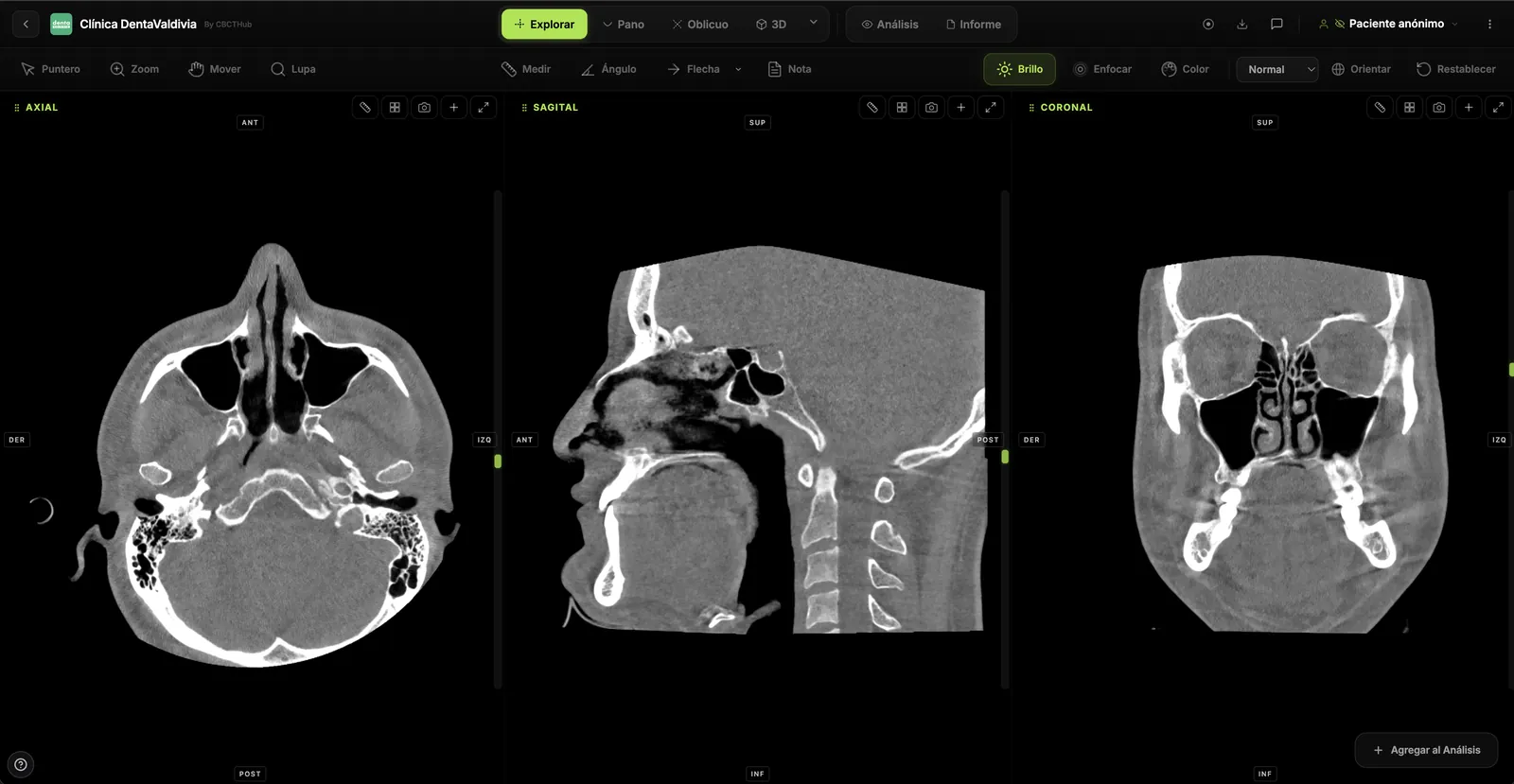
CBCT transverse analysis is the measurement, on coronal slices of the volume, of maxillary and mandibular basal width and of molar buccolingual inclination, in order to separate skeletal discrepancy from dentoalveolar compensation. Its clinical value lies in detecting maxillary deficiencies that casts and occlusion do not show: patients without crossbite can have a basal discrepancy masked by tipped molars[1].
Key points
- In a sample of 241 CBCT scans, Miner and colleagues found that many patients without crossbite had a skeletal transverse discrepancy compensated by molar inclination[1].
- For predicting crossbite, Miner's CBCT transverse analysis showed 87.0% sensitivity and 92.4% specificity, versus 70.4% and 57% for the J-point (jugale) analysis on simulated PA cephalograms[2].
- The systematic review by Sawchuk and colleagues concluded that diagnostic evidence on maxillary transverse deficiency is heterogeneous and of moderate to low quality, although CBCT appears more reliable than PA cephalometry[3].
- Dental arch widths can be the same in Class I and Class III while basal widths differ significantly: the dental level does not reveal the basal problem[4],[5].
- Dental measurements on CBCT reconstructions are highly reliable (r > 0.90), with a slight systematic underestimation that becomes significant when several measurements are combined[6].
- CBCT in orthodontics must be justified case by case, with FOV and exposure tailored to the clinical question, especially in children (ALADAIP principle)[7],[8].
Why perform a CBCT transverse analysis instead of relying on casts?
Traditional transverse diagnosis relies on occlusion, dental casts and arch-width indices. The problem is that these tools measure crowns, and crowns tip to compensate. The systematic review by Sawchuk and colleagues found arch-width prediction indices on casts to be unreliable for diagnosis, and frontal cephalograms derived from CBCT to be more reliable than conventional PA cephalograms for intermaxillary transverse assessment[3].
CBCT lets you measure basal bone and the molar axis on the same coronal slice. That separates two questions that casts conflate: is the maxilla narrow? and are the molars tipped? The answer determines whether the indication is skeletal expansion, dentoalveolar movement, or both.
For the broader orthodontic context, see CBCT in orthodontic treatment planning; this article focuses only on the transverse plane.
What does a CBCT transverse analysis measure?
There is no single standardized transverse analysis. Published methods share two groups of variables: skeletal basal widths and the dentoalveolar relationship (dental arch widths and molar inclination)[1],[4],[5].
Skeletal dimension: maxillary and mandibular basal width
In the study by Liu and colleagues, maxillary basal width was measured between the points where the tuberosity outline meets the zygomatic buttress, on the coronal slice of the first molars, and mandibular basal width between the most prominent points of the buccal alveolar process at the level of the mandibular first molars' center of resistance[5]. Koo and colleagues, at Yonsei University, measured basal width at each tooth's estimated center of resistance: the coronal third of the root in single-rooted teeth and the furcation in multirooted teeth[4].
Dentoalveolar dimension: molar buccolingual inclination
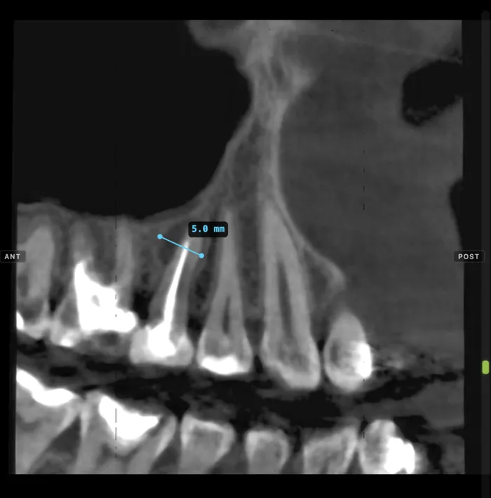
First molar inclination is measured between the tooth's long axis and a reference plane on the reoriented coronal slice. Diez-Rodrigálvarez and colleagues, in 40 subjects with normal occlusion, found mean angulations of about 99° for maxillary and 79° for mandibular molars relative to planes parallel to the occlusal plane, with no right-left differences and a negative correlation between arches that they interpret as dentoalveolar compensation[9]. These values depend on the reference method and should not be transferred to another protocol without adjustment.
| Variable | Where it is measured | What it indicates | Common error |
|---|---|---|---|
| Maxillary basal width | Coronal slice at first molars, at the zygomatic buttress/tuberosity[5] | Transverse size of the maxilla | Measuring on a slice tilted by head rotation |
| Mandibular basal width | Buccal alveolar process at the molar's center of resistance[5] | Reference against which the maxilla is compared | Picking points at different vertical levels on each side |
| Maxillomandibular basal difference | Subtraction of both basal widths[4],[5] | Skeletal discrepancy | Interpreting it without knowing the norm's method |
| Intermolar dental width | Central fossae or cusp tips of first molars[5] | Crown relationship | Assuming a normal width rules out basal deficiency |
| Molar buccolingual inclination | Molar long axis versus a reference plane[9] | Degree of dental compensation | Using norms from another reference system |
How do you tell skeletal discrepancy from dental compensation?
The key is to read basal width and molar inclination together. Miner and colleagues reported that the non-crossbite group included patients with normal transverse relationships and also a surprising number with an obvious skeletal discrepancy masked by dental compensation. In unilateral crossbite, the maxillary first molar on the non-crossbite side was compensated, whereas in bilateral crossbite molar inclinations were normal[1].
Liu and colleagues showed this in the early mixed dentition: in skeletal Class III children without crossbite, the maxillary-minus-mandibular basal width difference was −0.25 ± 1.73 mm versus 4.20 ± 1.25 mm in Class I, with no difference in dental arch widths. Maxillary molars were more buccally inclined (31.4° vs 17.64°) and mandibular molars more lingually inclined (45.24° vs 37.96°)[5]. In Class III adults, Koo and colleagues found the same pattern: similar dental widths and significantly different basal widths[4].
Clinical pearl: a “normal” transverse occlusion with markedly buccally tipped maxillary molars and lingually tipped mandibular molars is a compensation pattern. Expanding only at the dental level worsens the inclination that was already compensating.
Step-by-step transverse measurement protocol
- Reorient the volume. Align the midsagittal plane and the occlusal (or palatal, per your protocol) plane before measuring. Diez-Rodrigálvarez and colleagues used a standardized three-plane reorientation[9].
- Locate the coronal slice. Place it through the first molars; Miner and colleagues used a coronal cross-section through the middle of the maxillary and mandibular first molar crowns[2].
- Measure basal width of the maxilla and mandible with a defined method, always the same[4],[5].
- Measure the inclination of each first molar against the reference plane[9].
- Classify the finding with the algorithm and record the method in the report.
How reliable is CBCT transverse analysis?
Dental measurements on CBCT are reproducible. Baumgaertel and colleagues compared 10 measurements on 30 skulls, including intermolar and intercanine widths, against a digital caliper: reliability was high (r > 0.90) and CBCT slightly underestimated true anatomy, significantly only for compounded measurements[6]. Diagnostic validity is a separate matter.
| Method | Sensitivity | Specificity | PPV | NPV |
|---|---|---|---|---|
| CBCT transverse analysis (Miner) | 87.0% | 92.4% | 88.68% | 91.25% |
| J-point (jugale), simulated PA cephalogram | 70.4% | 57% | 52.78% | 73.77% |
| Transpalatal width measurement (TWM) | 98.1% | 13.9% | 43.8% | 91.67% |
Values for predicting clinical crossbite in 133 patients without dental compensation[2]. Predictive values depend on crossbite prevalence in the sample and do not transfer directly to other populations.
Common interpretation errors
- Measuring without reorienting: head rotation changes the distance between landmarks and the apparent molar inclination.
- Comparing with norms obtained by another method or at another age: Liu's values are from children aged 7 to 9[5]; Koo's, from adults[4].
- Reading molar inclination without basal width, or vice versa.
- Using a voxel size or FOV that does not clearly show the zygomatic buttress or both sides' molars. See also CBCT FOV selection.
What should the radiology report of a transverse analysis include?
The orthodontist needs figures that are comparable across follow-ups and an explicit method. From the imaging center, the useful deliverable is measurements and description, not a treatment recommendation.
| Finding | Meaning | What to report |
|---|---|---|
| Reduced or negative basal difference | Possible maxillary skeletal transverse deficiency | Basal widths in mm, method and slice level |
| Buccally tipped maxillary and lingually tipped mandibular molars | Dentoalveolar compensation | Inclination of each molar and reference plane |
| Asymmetric molar inclination | Unilateral compensation or asymmetry | Right and left values separately |
| Normal dental width with altered basal width | Masked discrepancy | Both widths, stating the dissociation |
If the FOV includes the TMJs or the airway, the radiologist must read the whole volume and describe findings outside the region of interest. For the joint, see what TMJ CBCT can assess.
When is CBCT justified for transverse assessment?
The American Academy of Oral and Maxillofacial Radiology (AAOMR) position statement on CBCT in orthodontics states that its use should be justified individually based on clinical presentation, with dose-optimization protocols[7]. In children, the European DIMITRA project proposes the ALADAIP principle (As Low As Diagnostically Acceptable, being Indication-oriented and Patient-specific)[8]. In practice, transverse analysis often uses a CBCT already indicated for another reason, or is justified when the expansion decision depends on separating skeletal from dental components.
How to do it in CBCTHub
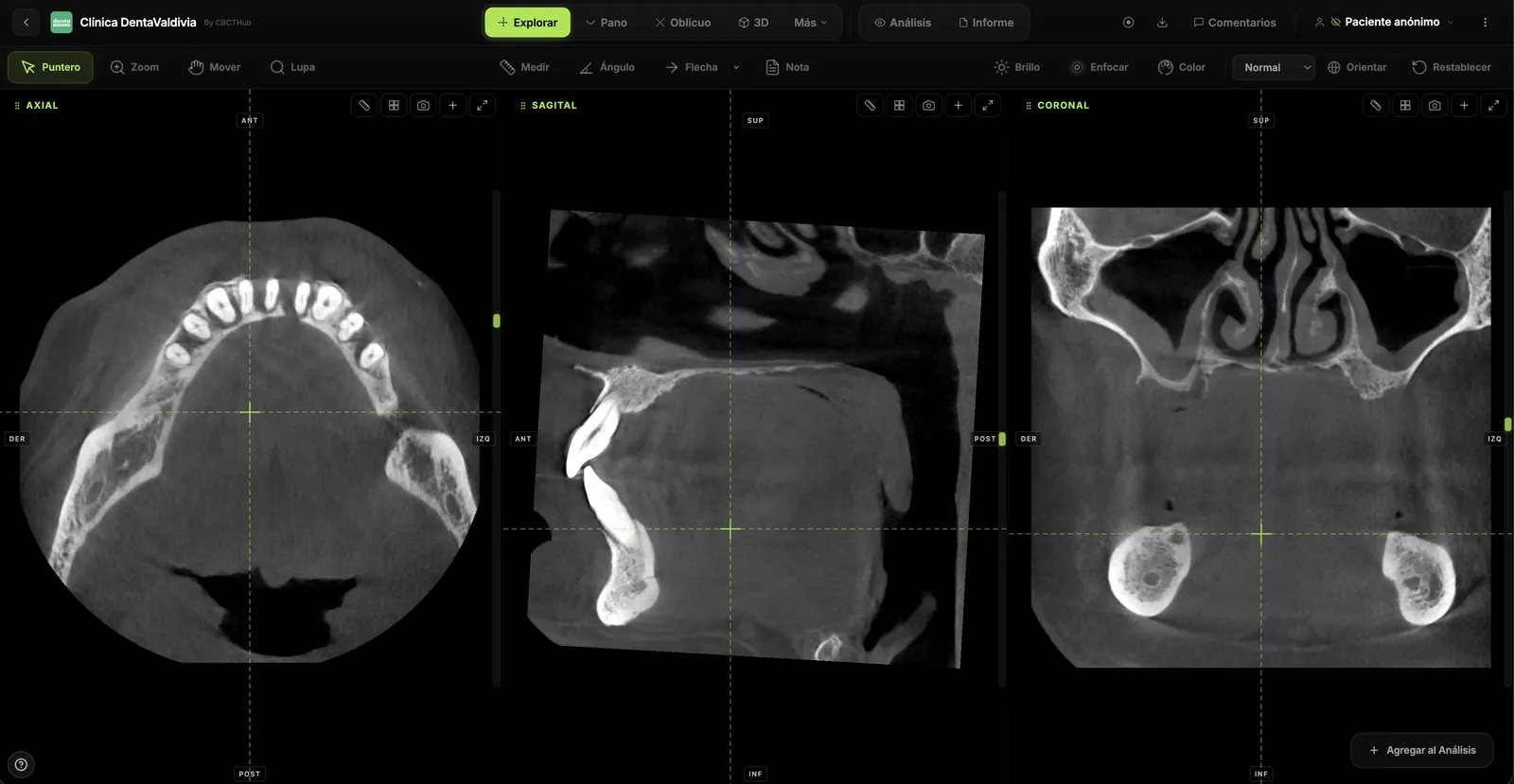
CBCTHub lets you perform the transverse analysis in the browser with no software to install; DICOM data is processed locally.
- Open the study from the link sent by the imaging center (or load the DICOM in the viewer).
- In the Explore (MPR) view, use Orient to align the midsagittal and occlusal planes.
- Place the coronal slice at the first molars; if the tooth axis does not line up, use an oblique slice.
- Use Measure for basal and dental widths in mm and Angle for each molar's inclination.
- Record the values in the report editor or leave slice-linked comments for the orthodontist.
For radiologists and imaging centers: deliver the study and report to referring dentists by link, no CDs, with your center's branding and PIN access. For orthodontists and dentists: open and measure the CBCT your imaging center sends you, right in the browser. Try the viewer free without an account at cbcthub.com/viewer or create an account at cbcthub.com.
Conclusion
CBCT transverse analysis provides what casts cannot: separating basal width from molar inclination. Evidence shows compensated skeletal discrepancies in patients without crossbite[1],[5], but also that methods are not standardized[3]. Always measure with the same protocol, report the method, and use the result to support, not replace, clinical judgment.
Frequently asked questions
What is a CBCT transverse analysis?
It is a set of measurements on coronal CBCT slices that compares maxillary with mandibular basal width and records the buccolingual inclination of the first molars. Its purpose is to determine whether a transverse discrepancy is skeletal, dentoalveolar or both, which casts cannot do because they only measure crowns that may already be compensated by tooth inclination.
Can a patient without crossbite have a maxillary transverse deficiency?
Yes. Miner and colleagues found, among 241 CBCT scans, patients without crossbite whose skeletal discrepancy was masked by dental compensation. Liu and colleagues observed in Class III children without crossbite a mean basal difference of −0.25 mm versus 4.20 mm in Class I, with similar dental widths and more buccally inclined maxillary molars.
On which slice is molar inclination measured?
On a coronal slice through the first molars, after reorienting the volume to the midsagittal plane and the occlusal or palatal plane. The angle is measured between the molar's long axis and the chosen reference plane. Because each study uses its own reference, normal values are comparable only when the same method is applied.
How reliable are transverse measurements on CBCT?
Dental measurements on CBCT, including intermolar widths, showed high reliability (r greater than 0.90) against a caliper on skulls, with a slight systematic underestimation. Reliability depends on reorienting the volume and using well-defined landmarks. According to Sawchuk's review, the diagnostic validity of transverse analyses still needs higher-quality studies.
Is CBCT justified only for transverse analysis?
The AAOMR states that CBCT in orthodontics should be justified individually based on clinical presentation, and the ALADAIP principle applies in children. Transverse analysis often uses a CBCT indicated for another reason, such as impacted teeth; it is justified on its own when the treatment decision depends on separating the skeletal from the dental component.
References
- Miner RM, Al Qabandi S, Rigali PH, Will LA. Cone-beam computed tomography transverse analysis. Part I: Normative data. Am J Orthod Dentofacial Orthop. 2012;142(3):300-7. doi:10.1016/j.ajodo.2012.04.014
- Miner RM, Al Qabandi S, Rigali PH, Will LA. Cone-beam computed tomography transverse analyses. Part 2: Measures of performance. Am J Orthod Dentofacial Orthop. 2015;148(2):253-63. doi:10.1016/j.ajodo.2015.03.027
- Sawchuk D, Currie K, Vich ML, Palomo JM, Flores-Mir C. Diagnostic methods for assessing maxillary skeletal and dental transverse deficiencies: A systematic review. Korean J Orthod. 2016;46(5):331-42. doi:10.4041/kjod.2016.46.5.331
- Koo YJ, Choi SH, Keum BT, Yu HS, Hwang CJ, Melsen B, Lee KJ. Maxillomandibular arch width differences at estimated centers of resistance: Comparison between normal occlusion and skeletal Class III malocclusion. Korean J Orthod. 2017;47(3):167-75. doi:10.4041/kjod.2017.47.3.167
- Liu F, Huang H, Shi X, Liu Y, Liu D. A study of transverse maxillomandibular discrepancy and dental compensation in early mixed dentition with skeletal Class III malocclusion without posterior crossbite. PLoS One. 2023;18(6):e0287343. doi:10.1371/journal.pone.0287343
- Baumgaertel S, Palomo JM, Palomo L, Hans MG. Reliability and accuracy of cone-beam computed tomography dental measurements. Am J Orthod Dentofacial Orthop. 2009;136(1):19-25. doi:10.1016/j.ajodo.2007.09.016
- American Academy of Oral and Maxillofacial Radiology. Clinical recommendations regarding use of cone beam computed tomography in orthodontics. Position statement by the American Academy of Oral and Maxillofacial Radiology. Oral Surg Oral Med Oral Pathol Oral Radiol. 2013;116(2):238-57. doi:10.1016/j.oooo.2013.06.002
- Oenning AC, Jacobs R, Pauwels R, Stratis A, Hedesiu M, Salmon B; DIMITRA Research Group. Cone-beam CT in paediatric dentistry: DIMITRA project position statement. Pediatr Radiol. 2018;48(3):308-16. doi:10.1007/s00247-017-4012-9
- Diez-Rodrigálvarez D, Bonilla-Morente E, López-Jiménez AJ. A Novel Method for the Diagnosis of Transverse Maxillary Deficiencies Based on CBCT. Diagnostics (Basel). 2026;16(7):1034. doi:10.3390/diagnostics16071034
Try CBCTHub for free
Upload, view, and share DICOM scans in the cloud. Nothing to install.
Create free accountRelated articles

TMJ CBCT: what it can assess and where it falls short
TMJ CBCT: which osseous changes it shows, what it misses compared with MRI, and how to report condylar findings using DC/TMD imaging criteria.

CBCT pre-implant assessment: what to check first
CBCT pre-implant assessment, step by step: cross-sectional measurements, the 2 mm safety margin, critical structures and what the report must include.

Endodontic CBCT: AAE/AAOMR 2025 indications
When to justify CBCT in endodontics per the AAE/AAOMR 2025 Position Statement: selective use, limited FOV, ALADAIP, and a practical reading checklist.