Using CBCT for Third Molar Extraction Planning

· Updated
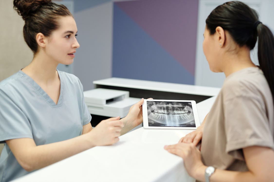
Why CBCT Matters for Wisdom Teeth
Third molar extraction is one of the most common surgical procedures in dentistry. While many cases are straightforward, a significant percentage involve close proximity to the inferior alveolar nerve (IAN), unusual root morphology, or proximity to the maxillary sinus. CBCT for third molar extraction provides the 3D information needed to plan a safe surgical approach.
Assessing Nerve Proximity
The most critical concern in lower third molar surgery is the relationship between the roots and the inferior alveolar nerve canal. On a panoramic radiograph, several signs suggest close proximity — darkening of the root at the canal crossing, deflection of the canal, or interruption of the white cortical line. However, these 2D signs cannot determine whether the nerve is buccal, lingual, or directly below the roots.
CBCT solves this by showing the exact spatial relationship in cross-section. The surgeon can see whether the nerve runs buccal to the roots, lingual, or directly inferior — each scenario requires a different surgical approach to minimize nerve injury risk.
Root Morphology Assessment
Curved, dilacerated, or hypercementosed roots significantly increase extraction difficulty. CBCT reveals:
- Number of roots: Supernumerary roots or fused roots that aren't visible on panoramic films
- Root curvature: The direction and degree of curvature in 3D, allowing the surgeon to plan the path of extraction
- Root tip proximity to vital structures: How close the apex is to the nerve canal, sinus floor, or adjacent tooth roots
Maxillary Third Molars and the Sinus
Upper wisdom teeth can have roots projecting into the maxillary sinus. CBCT shows the exact relationship between the root apices and the sinus floor, helping predict whether an oroantral communication is likely. If the sinus membrane is thin or absent over the roots, the surgeon can prepare for possible sinus exposure.
Surgical Approach Planning
With CBCT data, the surgeon can determine:
- Whether a buccal or lingual approach is safer based on nerve position
- How much bone removal is needed for access
- Whether sectioning the tooth is necessary and the optimal cut direction
- The presence of pathology (dentigerous cyst, pericoronitis extent) that might alter the surgical plan
Sharing the Surgical Plan
With online CBCT viewers, surgeons can share the scan with the referring dentist and the patient. Showing patients the 3D relationship between their wisdom tooth and the nerve helps them understand why surgery is recommended and what the risks are — improving informed consent and patient trust.
Try CBCTHub for free
Upload, view, and share DICOM scans in the cloud. Nothing to install.
Create free accountRelated articles
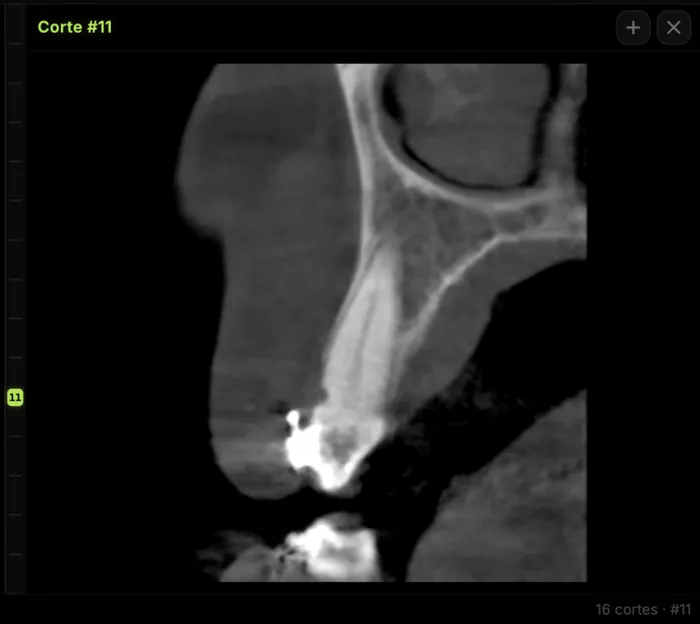
Mandibular canal and inferior alveolar nerve on CBCT: anatomy, variants, and pre-surgical measurement
Locate the mandibular canal and IAN on CBCT: mental loop, bifid/retromolar canal, measurement checklist, and when to escalate from panoramic (ALADA).
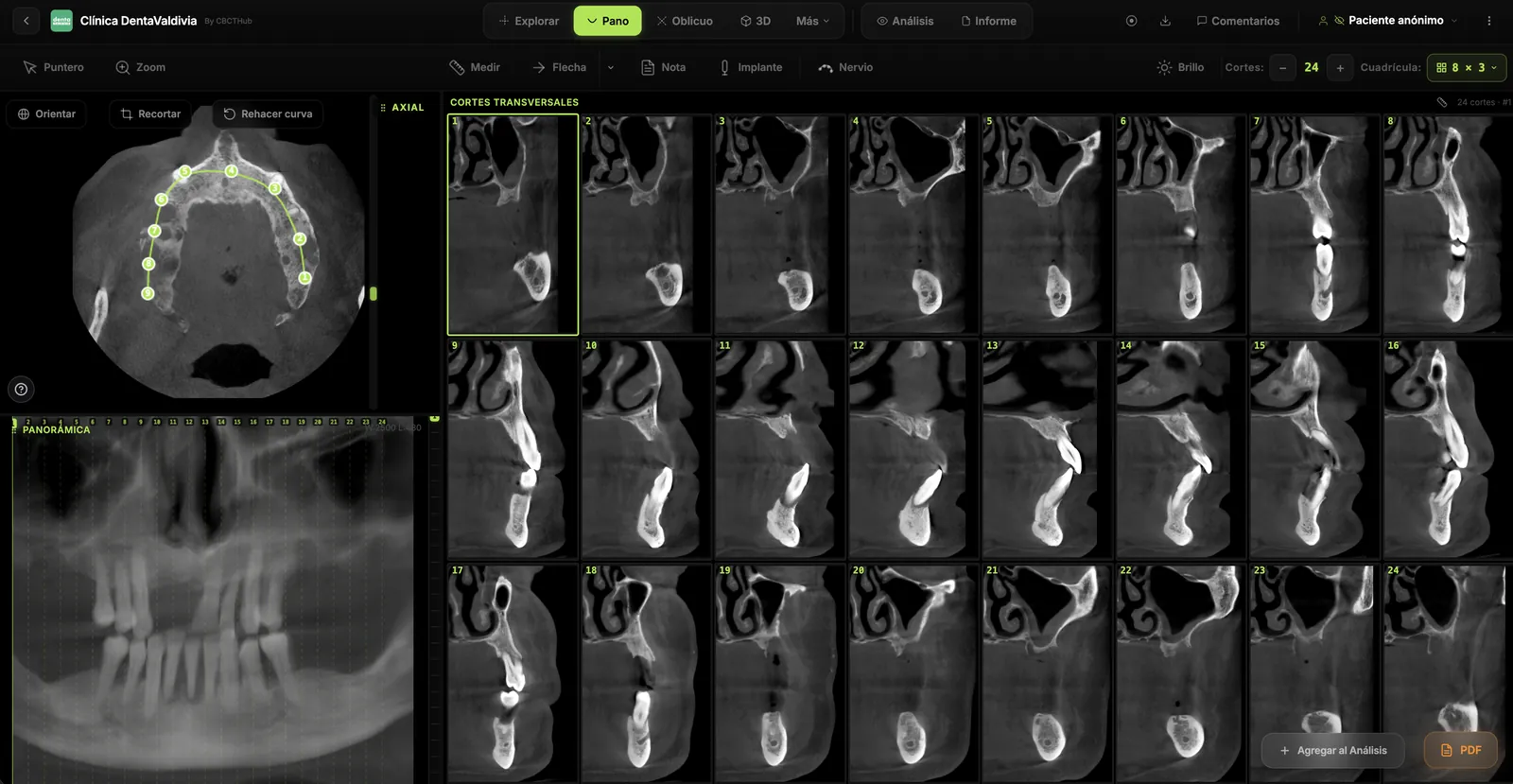
Browser DICOM viewer vs desktop: what works for CBCT in clinic
Open CBCT in the browser or install a desktop viewer? A clinical decision matrix: MPR performance, offline use, Mac/Windows mix, sharing without CDs, and when native software still wins.
How to open a CBCT without installing software: a practical online-viewer guide
You got a CD, ZIP, or link with a CBCT and no manufacturer viewer. How to open the DICOM in the browser, measure in mm, and what to do when it fails.