Mandibular canal and inferior alveolar nerve on CBCT: anatomy, variants, and pre-surgical measurement
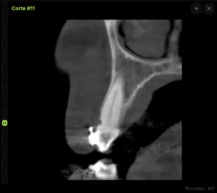
The mandibular canal and the inferior alveolar nerve (IAN) are the most critical anatomical limits in implantology, third-molar surgery, and regional anesthesia. On panoramic radiography their course is projected in 2D, and variants (bifid canal, retromolar canal, accessory foramina) are underestimated. CBCT lets you trace the canal in three planes, measure real distances to the crest and to the lingual cortex, and decide whether the risk justifies changing the surgical plan.
Why a panoramic is not enough
The panoramic remains an excellent screening tool: it shows the full mandibular arch at low dose. But it distorts vertically, superimposes structures, and does not reliably measure crestal–canal distance or the buccolingual position of the IAN. When the clinical plan hinges on millimeters (posterior implant, osteotomy near the mental foramen, third molar in intimate contact with the canal), the evidence and selective-use CBCT guidelines agree: escalate to 3D only if the result can change management—not routinely.
The applicable principle is ALADA / ALADAIP: As Low As Diagnostically Acceptable, indication- and patient-specific. The joint AAE–AAOMR 2025 update on CBCT in endodontics reinforces the same message for dentistry in general: selective CBCT, individualized protocols, and training in interpretation—not an “always 3D” protocol.
Anatomy you must locate in the volume
Main mandibular canal
It runs through the mandibular body from the mandibular foramen to the mental foramen, where the nerve divides into the mental nerve and, frequently, an anterior loop. On CBCT it is good practice to:
- Identify the canal on axial and coronal views (cross-sections perpendicular to the panoramic curve).
- Follow the course on oblique sagittal slices or along the panoramic reconstruction of the volume.
- Measure crestal–canal roof distance at the planned surgical site, not on a “nice” slice away from the area of interest.
Anterior loop of the mental nerve
The anterior loop can extend several millimeters mesial to the mental foramen. When planning implants in the premolar region, seeing the foramen is not enough: you must measure loop length on the corresponding axial/coronal plane. An inadequate safety margin here explains neurosensory injuries despite a CBCT that was only “eyeballed.”
Variants: bifid canal, retromolar canal, and accessory foramina
CBCT studies report accessory / bifid canals with variable prevalence depending on population and radiographic criteria; European series have described variants in a substantial proportion of patients when sought systematically. A recent study measured bifid-canal frequency near 9% in a CBCT cohort, with mean diameters on the order of 1–2.5 mm (larger in the retromolar region). Not every visible “double canal” is clinically relevant, but they matter when:
- There is repeated failure of IAN block with soft-tissue anesthesia present and residual dental sensitivity (an accessory branch not reached by the conventional technique).
- Osteotomy, third-molar extraction, or implant placement is planned along the course of a retromolar or bifid canal.
- Unexplained paresthesia or bleeding occurs after surgery in a zone that looked “safe” on panoramic.
A clinical-management review of mandibular canal variants proposes a PR → selective CBCT → MRI-only-in-selected-cases pathway, with clear triggers: equivocal panoramic, short crestal–canal distance (e.g. ≤4–5 mm at the site), suspected variant, complex third molars, repeated IANB failures, or persistent neurosensory symptoms.
How to measure in practice (reading checklist)
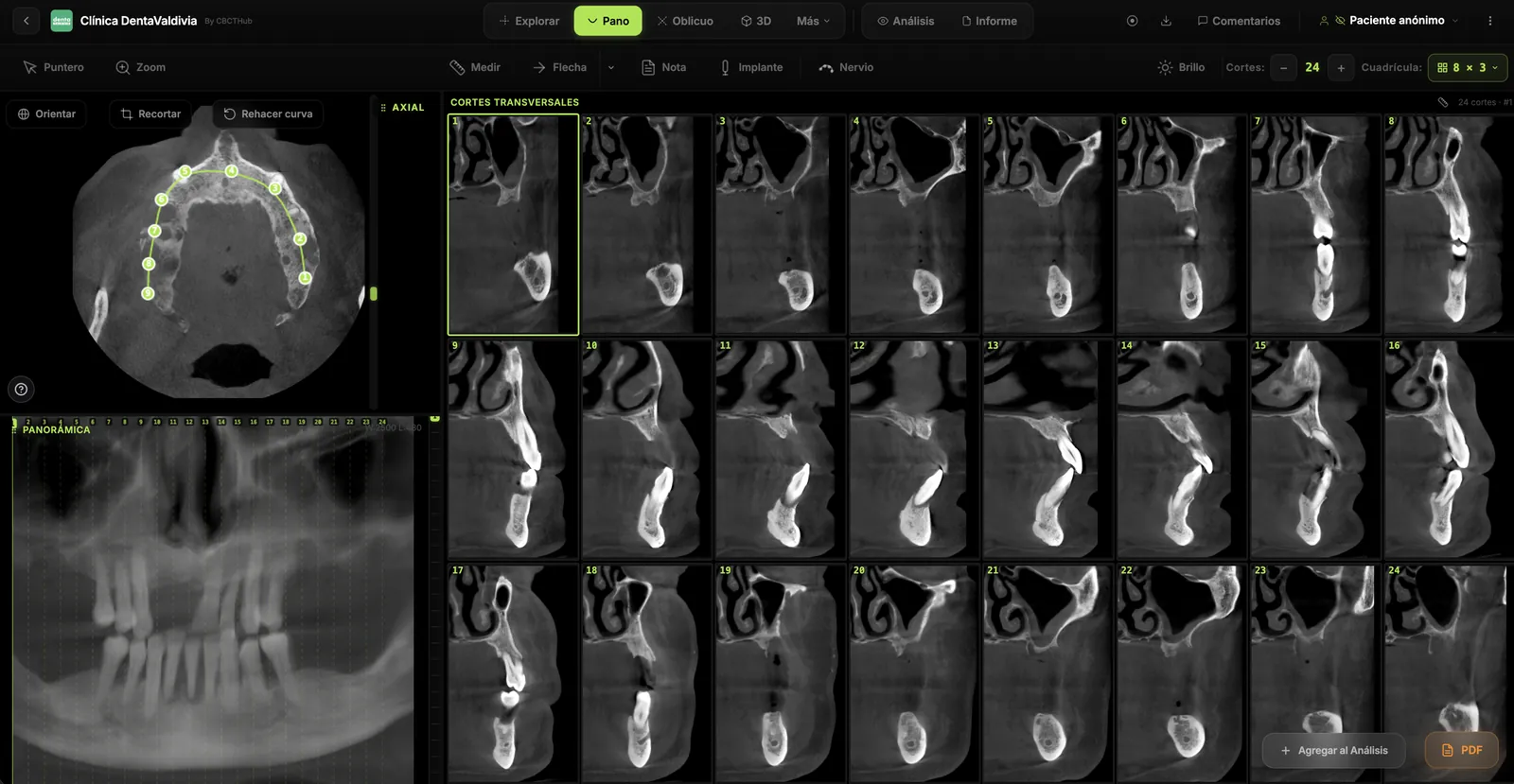
- Orient the volume to the occlusal plane and mandibular arch; generate the panoramic curve and cross-sections at the site of interest.
- Mark the canal roof on every relevant slice; measure crestal–canal mm and distance to lingual/buccal cortex.
- Locate the mental foramen and the loop in the premolar zone before osteotomies or implants there.
- Hunt for variants: double cortex, retromolar course toward the trigone, accessory foramina on the buccal surface.
- Document in the report: side, distances, presence/absence of variants, and recommendation (safety margin, alternative anesthetic technique, need for a surgical pilot drill, etc.).
Voxel size matters: for IAN–root relationships in third molars or fine detail, a voxel ≤0.2 mm is usually more useful than a large FOV with a coarse voxel. Choose the smallest FOV that answers the clinical question (ALARA/ALADA).
Clinical implications by scenario
Posterior implants
Crestal–canal distance defines maximum implant length and the need for augmentation or more coronal positioning. A lingual canal position changes the risk of lingual perforation and floor-of-mouth bleeding. Measure along the planned implant axis, not only along the axis of the missing tooth.
Third molars
Intimate contact, interruption of the canal cortex, root darkening, or canal deviation on panoramic are classic signals to request CBCT. 3D clarifies whether the root is buccal, lingual, or interlaced with the IAN and helps choose coronectomy versus complete extraction.
Anesthesia
If the patient has a history of incomplete blocks, look for a bifid/retromolar canal. Higher techniques (e.g. Gow-Gates) or adjuncts (Vazirani-Akinosi, infiltrations, periodontal ligament) may be needed when an accessory branch escapes the conventional block.
Common errors when reading the IAN on CBCT
- Relying on a single “representative” slice instead of scrolling the full surgical segment.
- Ignoring the anterior loop in the premolar region.
- Treating relative CBCT densities as absolute Hounsfield units when describing canal “content.”
- Using a large maxillofacial FOV with a coarse voxel for a millimeter-level question in one quadrant.
- Failing to correlate with the clinic (symptoms, anesthesia failures, surgical plan).
FAQ
Should I order CBCT for every patient with a mandibular implant?
Not routinely. It is indicated when panoramic or clinical findings leave a doubt that 3D can resolve (short distance, doubtful foramen/loop, suspected variant, complex anatomy). Benefit must outweigh the additional dose.
Does CBCT replace the panoramic?
No. They are complementary: panoramic for screening and the full arch; CBCT for specific spatial questions.
What should go in the radiology report?
Canal course, distances at the site of interest, mental foramen and loop if applicable, variants, and the clinical implication in one sentence (margin, risk, suggested technique).
References
- American Association of Endodontists; American Academy of Oral and Maxillofacial Radiology. Use of Cone-Beam Computed Tomography in Endodontics — 2025 Update (joint position statement). AAE Newsroom / societies.
- Imaging-Based Clinical Management of Mandibular Canal Variants: PR–CBCT–Selective MRI. Biomedicines. 2025. DOI: 10.3390/biomedicines13112760
- Bifid mandibular canal assessment and its relevance to local anesthesia. J Dent Anesth Pain Med. 2025;25(3):183. DOI: 10.17245/jdapm.2025.25.3.183
- Prevalence and Characteristics of Accessory Mandibular Canals: A Cone-Beam Computed Tomography Study in a European Adult Population. Diagnostics. 2022;12(8):1885. DOI: 10.3390/diagnostics12081885
Note: this article is educational and does not replace clinical judgment or formal radiographic reading of the individual case.
Try CBCTHub for free
Upload, view, and share DICOM scans in the cloud. Nothing to install.
Create free accountRelated articles
How to open a CBCT without installing software: a practical online-viewer guide
You got a CD, ZIP, or link with a CBCT and no manufacturer viewer. How to open the DICOM in the browser, measure in mm, and what to do when it fails.
Online CBCT viewer: what it is and when to use it in clinic
What an online CBCT viewer is, when to use it vs desktop or CD, DICOM tools (MPR, measurements, share link) and basic privacy in clinic.
Best CBCT Viewers 2026: Complete Comparison of 10 Dental Imaging Software Options
In-depth comparison of the best CBCT viewers for dental imaging in 2026. Cloud-based and desktop options, pricing, features, and best-fit scenarios.