CBCT indications in dentistry: when to order and when not

CBCT indications in dentistry come down to one rule: a scan is indicated when there is a specific clinical question, history and clinical examination have been completed, lower-dose conventional radiography cannot answer it, and the answer could change the diagnosis or treatment plan [1][2]. CBCT is not indicated for routine or screening use, caries detection, routine assessment of periodontal bone support, or questions about soft tissues [2].
This article organizes European and North American CBCT indications in dentistry by clinical area, reviews what the evidence says about the real impact of a volume on decisions, and outlines what a well-justified referral should contain and what responsibility the reader of the scan takes on.
Key points
- No CBCT should be requested without a prior history and clinical examination; "routine" or screening use is considered unacceptable practice [2].
- CBCT is justified only when lower-dose conventional radiography cannot answer the question [1].
- The ADA advises prescribing CBCT only when the expected diagnostic yield will benefit patient care, enhance patient safety or improve clinical outcomes significantly [3].
- Effective dose varies widely across units and protocols: published adult values range from 5 to 1073 µSv [4].
- A well-indicated CBCT changes decisions: in endodontics it altered the treatment plan in 53% of patients selected with European criteria [5].
- Whoever reads the scan must evaluate the entire acquired volume, not just the region of interest [1].
What does CBCT justification mean?
CBCT justification means demonstrating, for this patient and this question, that the expected diagnostic benefit outweighs the risk of exposure. The SEDENTEXCT guidelines, published by the European Commission as Radiation Protection No 172, require justification to be individual, the scan to be able to add new information to patient management, and the justification to be recorded in the clinical notes [2]. The 20 basic principles of the European Academy of Dental and Maxillofacial Radiology (EADMFR) add that CBCT should be used only when lower-dose conventional radiography cannot answer the question, and that the smallest volume compatible with the clinical situation should be selected if it lowers the dose [1].
An often overlooked point: the referring dentist must provide enough clinical information for the practitioner performing the scan to justify it [2]. A request that only says "maxillary CBCT" gives no basis for choosing FOV, voxel size or protocol, and shifts the decision to the imaging center without data.
CBCT indications in dentistry by clinical area
A review of 26 publications containing CBCT guidance found broad agreement on clinical use, except for treatment planning in implant dentistry, and frequently low methodological quality when appraised with the AGREE II instrument [6]. The table summarizes when to order a CBCT and when not to, according to the most cited sources.
| Area | When to order | When not / alternative |
|---|---|---|
| Caries | — | Not indicated for caries detection or diagnosis [2] |
| Periodontics | Limited-volume, high-resolution CBCT in selected infrabony defects and furcation lesions [2] | Not as a routine method to image periodontal bone support [2] |
| Endodontics | Limited FOV when intraoral images are equivocal or inadequate: complex anatomy, resorption, retreatment, apical surgery [2][7][8] | Not as a standard method to identify periapical pathosis or show canal anatomy [2] |
| Implants | Cross-sectional imaging of all sites; CBCT is the imaging method of choice to obtain it [9] | Requests with no defined site or prosthetic plan |
| Mandibular third molar | When 2D imaging suggests a direct relationship with the mandibular canal and removal has been decided [2][10] | Periapical or panoramic imaging is sufficient in most cases [10] |
| Orthodontics | Localized assessment of impacted teeth with limited FOV; complex skeletal cases [2] | No routine large-FOV CBCT for orthodontic diagnosis; justify case by case [2][11] |
| TMJ | Osseous assessment; alternative to medical CT [2] | Disc and soft tissues: MRI [1] |
| Pathology and trauma | Osseous component of lesions, maxillofacial fractures, orthognathic planning when bone information is required [2] | When soft-tissue evaluation is needed, medical CT or MRI [2] |
For specialty-level detail, see our articles on endodontic CBCT indications in complex cases, CBCT pre-implant assessment and mandibular third molars and the IAN canal. For the basic comparison of both modalities, read CBCT vs panoramic radiograph; here the focus is on the justification reasoning.
When should you not order a CBCT?
CBCT is not indicated in these situations, even when the unit is available in the office:
- Screening or a "full check-up": routine use without history and clinical examination is unacceptable under SEDENTEXCT [2].
- Caries and routine periodontal monitoring: the evidence does not support it [2].
- Questions a periapical or panoramic already answers: the EADMFR principle is to use the lower-dose technique first [1].
- Soft-tissue questions: articular disc, soft-tissue extent of a lesion or tumors require MRI or medical CT [1][2].
- Large FOV "just in case": the field of view should match the question, not the maximum the unit allows [1].
Common mistake: requesting the largest FOV "to avoid a retake". If the question is a premolar with doubtful anatomy, a full-skull volume raises the dose, lowers useful resolution and forces the reader to report structures nobody needed to see.
How much does CBCT change clinical decisions?
Justification rests on diagnostic and therapeutic efficacy, not on image sharpness. Matzen and Wenzel assessed mandibular third molar imaging with the Fryback and Thornbury hierarchical model: most studies sit at the lower levels (technical capability and diagnostic accuracy), and very few address impact on treatment and patient outcome [10].
The multicentre randomised controlled trial by Ghaeminia et al. illustrates the point. In 268 patients with 320 high-risk third molars on panoramic imaging, the incidence of inferior alveolar nerve sensory disturbance at one week was 6.3%, with no significant difference between the CBCT and panoramic-only arms (p = 0.64) [12]. The authors concluded that CBCT identifies risk better but does not reduce nerve injury if the surgical technique stays the same; its value lies in choosing alternatives such as coronectomy [12].
In endodontics, the picture differs when selection is strict. In two specialist clinics, only 4% of referred patients met European criteria for CBCT; in that group the treatment plan changed in 53% of cases [5].
Clinical pearl: before ordering, ask what you would do differently depending on the result. If the answer is "nothing", the CBCT is not justified.
Dose, field of view and the ALADAIP principle
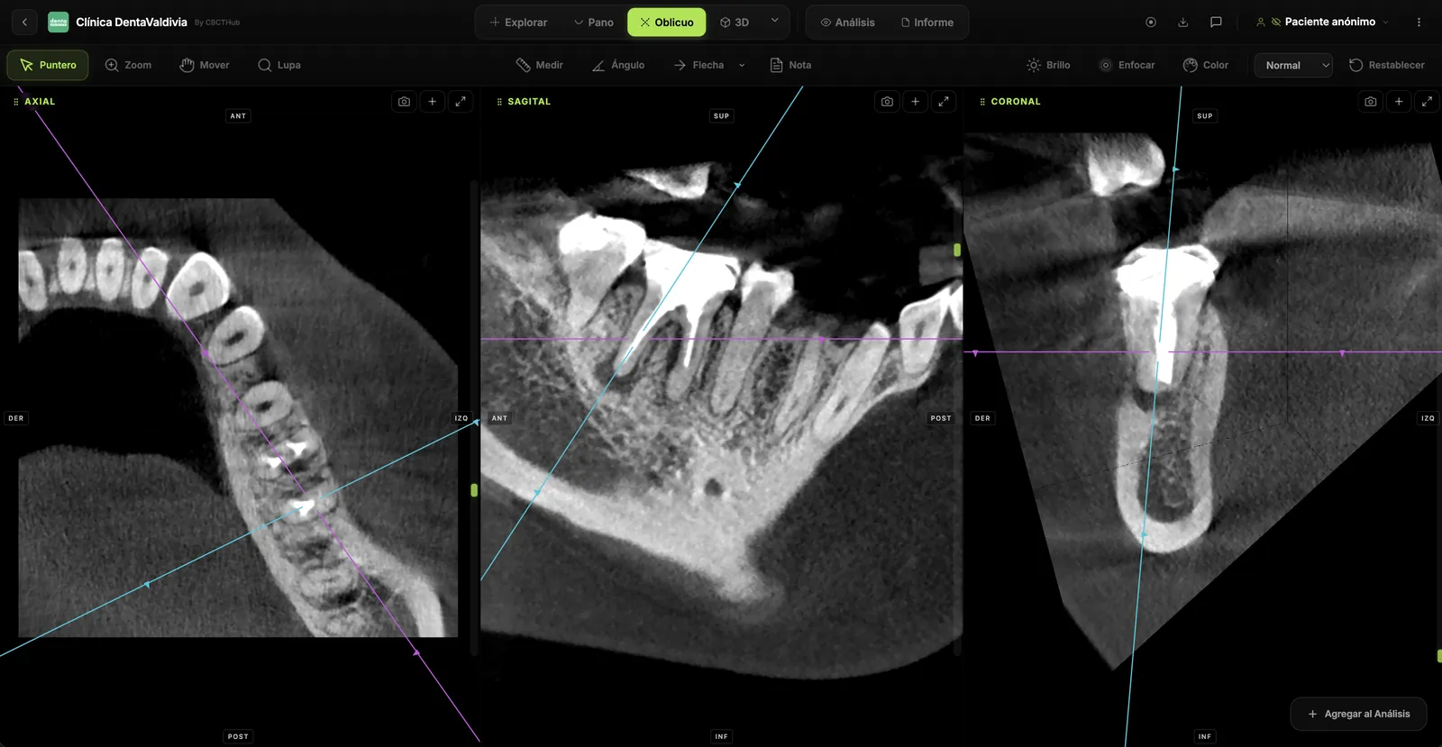
CBCT dose cannot be generalized. The meta-analysis by Ludlow et al. reported adult effective doses of 46 to 1073 µSv for large FOVs, 9 to 560 µSv for medium FOVs and 5 to 652 µSv for small FOVs, depending on unit and protocol [4]. Jacobs et al. put it in clinical terms: for similar indications, the dose may equal anywhere from 2 to 200 panoramic radiographs [13].
A small FOV reaching 652 µSv shows that field size alone is not enough: voxel size, mA, rotation and high-resolution modes also matter. Optimization has therefore evolved from ALARA and ALADA to ALADAIP (As Low As Diagnostically Acceptable, being Indication-oriented and Patient-specific) [14]. The European DIMITRA project applied this approach to children and adolescents, with indication-oriented and patient-specific recommendations [15].
What should a well-justified CBCT referral include?
| Element | Why it matters |
|---|---|
| Specific clinical question | Defines FOV, voxel size and protocol |
| Relevant clinical findings | Allows the imaging center to justify the exposure [2] |
| Previous radiographs and their findings | Avoids repeats and confirms 2D is insufficient [1] |
| Region and teeth of interest | Allows the smallest possible volume [1] |
| Decision that depends on the result | Shows the scan can change management [3] |
| Patient age and conditions | Protocol adjustment under ALADAIP [14] |
The role of the radiologist and the imaging center
Justification is shared. When the referral does not support it, the imaging center can propose a lower-dose technique or a smaller FOV and document it. Once acquired, the volume must be read in full: EADMFR principle 7 requires a thorough evaluation of the entire image dataset, not just the region that prompted the scan [1]. SEDENTEXCT adds that dentoalveolar volumes may be reported by an oral and maxillofacial radiologist or an adequately trained general dentist, whereas craniofacial volumes require an oral and maxillofacial radiologist or a clinical radiologist [2].
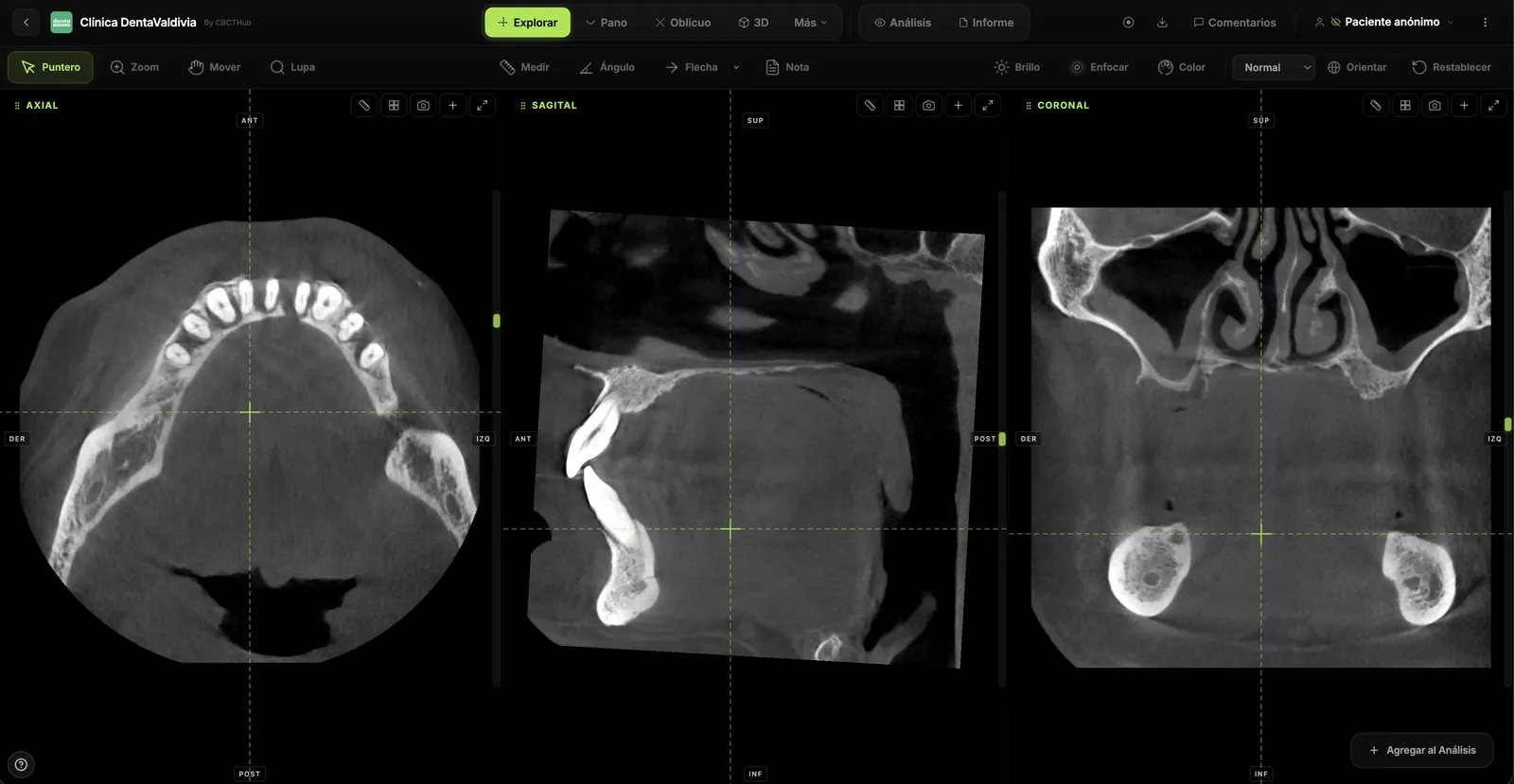
In practice, the report should state the indication received and the FOV and voxel used, answer the clinical question first, and then list findings in the rest of the volume with their relevance.
How to do it in CBCTHub
CBCTHub helps close the loop between indication, reading and communicating the result:
- Open the scan in the web viewer: DICOM is processed locally in the browser with no software to install, and it works with Carestream, Planmeca, Vatech, Sirona, Morita, NewTom and other units.
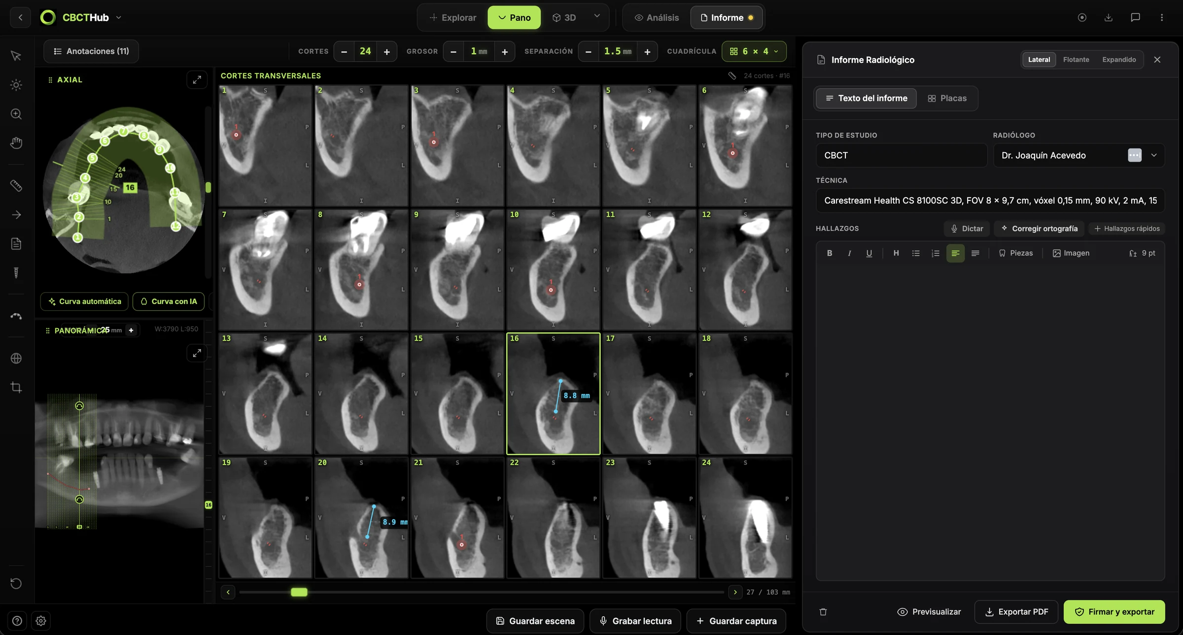
- Review the whole volume in the MPR view (axial, sagittal and coronal), and use oblique slices, panoramic with cross-sections, or the endodontic and TMJ modules depending on the question.
- Measure in millimeters and leave comments linked to the exact slice where the finding is.
- Write the report in the editor, including indication and FOV, and build PDF image plates.
- Share the scan and report by link, with a PIN for the referring dentist.
If you are a radiologist or run an imaging center, deliver scans and reports to your referring dentists by link, with no CDs. If you are a dentist or specialist, open and measure the CBCT your imaging center sends you right in the browser. Try the viewer free with no account at cbcthub.com/viewer or create an account at cbcthub.com.
Conclusion
Ordering a CBCT is a clinical decision, not a formality. A specific question, a prior clinical examination, the inadequacy of 2D imaging and a real chance of changing the plan are the four filters the guidelines rely on. When they are met, the volume should be acquired with the smallest useful FOV, read in full and reported clearly. This content is intended for professionals and does not replace case-by-case clinical judgment.
Frequently asked questions
When is a CBCT indicated in dentistry?
A CBCT is indicated when there is a specific clinical question, history and clinical examination are complete, lower-dose conventional radiography cannot answer it, and the result could change the diagnosis or treatment. This is set out in the EADMFR basic principles and the European Commission SEDENTEXCT guidelines. Justification must be individual and recorded in the patient's clinical notes.
Can CBCT be used for caries detection or periodontal monitoring?
Not routinely. The SEDENTEXCT guidelines state that CBCT is not indicated for caries detection or diagnosis, nor as a routine method to image periodontal bone support. Only in selected cases of infrabony defects or furcation lesions may a limited-volume, high-resolution CBCT be justified, when intraoral radiographs do not provide the information needed for the periodontal treatment decision.
Which field of view should be requested for a dental CBCT?
Request the smallest field of view compatible with the clinical question, provided it lowers the dose. A single tooth or implant site rarely needs a full-arch or skull volume. Field size alone does not define dose, though: the Ludlow meta-analysis found small-FOV adult doses as high as 652 µSv, so voxel size and protocol must also be tailored to the indication.
Who is responsible for justifying a CBCT scan?
Justification is shared. The referring dentist must provide the clinical question, examination findings and previous radiographs; the practitioner performing the scan must confirm the exposure is justified and choose the protocol. If the referral does not support it, the imaging center can propose a lower-dose technique. SEDENTEXCT requires that this justification be recorded in the patient's clinical records.
Must the whole volume be reported even if the question is localized?
Yes. The EADMFR basic principles require a thorough evaluation of the entire acquired dataset, not just the region that prompted the scan. The report should therefore answer the clinical question first and then describe findings in the rest of the volume, such as the maxillary sinuses, airway or bony structures, stating their relevance and whether follow-up or referral is needed.
References
- Horner K, Islam M, Flygare L, Tsiklakis K, Whaites E. Basic principles for use of dental cone beam computed tomography: consensus guidelines of the European Academy of Dental and Maxillofacial Radiology. Dentomaxillofac Radiol. 2009;38(4):187-95. doi:10.1259/dmfr/74941012
- European Commission. Radiation Protection No 172. Cone beam CT for dental and maxillofacial radiology: evidence-based guidelines (SEDENTEXCT). Luxembourg: Directorate-General for Energy; 2012. energy.ec.europa.eu
- American Dental Association Council on Scientific Affairs. The use of cone-beam computed tomography in dentistry: an advisory statement from the American Dental Association Council on Scientific Affairs. J Am Dent Assoc. 2012;143(8):899-902. doi:10.14219/jada.archive.2012.0295
- Ludlow JB, Timothy R, Walker C, Hunter R, Benavides E, Samuelson DB, et al. Effective dose of dental CBCT: a meta analysis of published data and additional data for nine CBCT units. Dentomaxillofac Radiol. 2015;44(1):20140197. doi:10.1259/dmfr.20140197
- Mota de Almeida FJ, Knutsson K, Flygare L. The effect of cone beam CT (CBCT) on therapeutic decision-making in endodontics. Dentomaxillofac Radiol. 2014;43(4):20130137. doi:10.1259/dmfr.20130137
- Horner K, O'Malley L, Taylor K, Glenny AM. Guidelines for clinical use of CBCT: a review. Dentomaxillofac Radiol. 2015;44(1):20140225. doi:10.1259/dmfr.20140225
- AAE and AAOMR Joint Position Statement: Use of Cone Beam Computed Tomography in Endodontics 2015 Update. J Endod. 2015;41(9):1393-6. doi:10.1016/j.joen.2015.07.013
- Sousa Melo SL, Fayad MI, Gohel A, Johnson BR, Kalathingal S, Mahdian M, et al. AAE and AAOMR Joint Position Statement: Use of Cone-Beam Computed Tomography in Endodontics 2025 Update. J Endod. 2026;52(1):4-13. doi:10.1016/j.joen.2025.09.008
- Tyndall DA, Price JB, Tetradis S, Ganz SD, Hildebolt C, Scarfe WC; American Academy of Oral and Maxillofacial Radiology. Position statement of the American Academy of Oral and Maxillofacial Radiology on selection criteria for the use of radiology in dental implantology with emphasis on cone beam computed tomography. Oral Surg Oral Med Oral Pathol Oral Radiol. 2012;113(6):817-26. doi:10.1016/j.oooo.2012.03.005
- Matzen LH, Wenzel A. Efficacy of CBCT for assessment of impacted mandibular third molars: a review - based on a hierarchical model of evidence. Dentomaxillofac Radiol. 2015;44(1):20140189. doi:10.1259/dmfr.20140189
- American Academy of Oral and Maxillofacial Radiology. Clinical recommendations regarding use of cone beam computed tomography in orthodontics. Position statement by the American Academy of Oral and Maxillofacial Radiology. Oral Surg Oral Med Oral Pathol Oral Radiol. 2013;116(2):238-57. doi:10.1016/j.oooo.2013.06.002
- Ghaeminia H, Gerlach NL, Hoppenreijs TJ, Kicken M, Dings JP, Borstlap WA, et al. Clinical relevance of cone beam computed tomography in mandibular third molar removal: a multicentre, randomised, controlled trial. J Craniomaxillofac Surg. 2015;43(10):2158-67. doi:10.1016/j.jcms.2015.10.009
- Jacobs R, Salmon B, Codari M, Hassan B, Bornstein MM. Cone beam computed tomography in implant dentistry: recommendations for clinical use. BMC Oral Health. 2018;18(1):88. doi:10.1186/s12903-018-0523-5
- Mendonça RP, Estrela C, Bueno MR, Carvalho TCASG, Estrela LRA, Chilvarquer I. Principles of radiological protection and application of ALARA, ALADA, and ALADAIP: a critical review. Braz Oral Res. 2025;39:e14. doi:10.1590/1807-3107bor-2025.vol39.014
- Oenning AC, Jacobs R, Pauwels R, Stratis A, Hedesiu M, Salmon B; DIMITRA Research Group. Cone-beam CT in paediatric dentistry: DIMITRA project position statement. Pediatr Radiol. 2018;48(3):308-16. doi:10.1007/s00247-017-4012-9
Try CBCTHub for free
Upload, view, and share DICOM scans in the cloud. Nothing to install.
Create free accountRelated articles

Mandibular Third Molar CBCT: Reading the IAN Canal
Mandibular third molar CBCT: when to order it, what to assess on cross-sections and how findings guide removal vs coronectomy. Evidence-based guide.

Endodontic CBCT: AAE/AAOMR 2025 indications
When to justify CBCT in endodontics per the AAE/AAOMR 2025 Position Statement: selective use, limited FOV, ALADAIP, and a practical reading checklist.

Endodontic CBCT indications: a guide to complex cases
Endodontic CBCT indications per AAE/AAOMR: which complex cases justify a scan, which FOV to use, what to report and where CBCT falls short. For clinicians.