Endodontic CBCT: AAE/AAOMR 2025 indications
CBCT in endodontics is not a routine screening exam. It is justified when the clinical examination and well-made periapical radiographs (or a panoramic when relevant) do not answer the diagnostic or treatment question. The AAE and AAOMR Joint Position Statement: Use of Cone-Beam Computed Tomography in Endodontics 2025 Update replaces the 2015 document, condenses guidance into 12 recommendations, and reinforces selective use, interpretation training, and the ALADAIP principle.
This article turns that framework into chairside decisions for dentists and endodontists: when 2D is enough, when to order a limited-FOV volume, how to request the protocol, and how to read it—without inventing dose figures or miracle cases. It is not a product pitch, not a full mandibular-canal anatomy review (IAN proximity for apical surgery is mentioned only briefly, with a link to the dedicated post), and not legal advice.
What the 2025 Update changes vs 2015
The 2015 AAE/AAOMR joint statement already framed CBCT as case-by-case, not screening. The 2025 Update—published in the Journal of Endodontics and in Oral Surgery, Oral Medicine, Oral Pathology and Oral Radiology in early 2026—keeps that logic and makes it more usable:
- It moves from 14 to 12 condensed recommendations spanning diagnosis, treatment, and outcome assessment.
- It reiterates that CBCT should be used selectively, guided by clinical indications, existing imaging, and the patient’s needs.
- It foregrounds ALADAIP: As Low As Diagnostically Acceptable, Indication- and Patient-specific.
- It places greater weight on training in CBCT use and interpretation for dentists and auxiliary staff, and on cross-specialty collaboration.
- It notes that artificial intelligence may support interpretation: an emerging aid, not a substitute for clinical judgment, and not a claim about any specific commercial viewer feature.
The AAE newsroom release (14 January 2026) also states that the guidance aligns with recent ADA/AAOMR findings that lead aprons and thyroid collars are no longer generally recommended for CBCT, while acknowledging jurisdiction-specific requirements. That is not permission to ignore local rules: follow what applies in your state or country.
In Europe, SEDENTEXCT (Radiation Protection No 172) and the European Society of Endodontology (ESE) 2014 position statement point the same way: justification + optimization + limited FOV for endodontic questions.
ALADAIP in clinic: from acronym to order form
ALADAIP does not replace ALARA; it specializes it. In endodontic practice it means:
- Write the indication. What question will the volume answer that a well-angulated periapical does not?
- Review prior imaging. If 2D already shows the missed canal, the obvious fracture pattern, or a lesion with clear borders for your plan, do not “complete the chart” with an automatic CBCT.
- Match the protocol. Small FOV centered on the tooth (or the smallest field that answers the question); high spatial resolution / small voxel when fine canal detail is required; immobilization to reduce motion; MAR when the system offers it and metal justifies the attempt.
- Patient-specific factors. Age, pregnancy (defer when diagnosis is not urgent), motion risk, and whether a recent useful study already exists.
Ordering a “large FOV just in case” violates ALADAIP. Ordering a limited FOV with the clinical question on the requisition fulfills it.
When 2D radiographs are enough
Quality periapicals remain first-line for most uncomplicated root canal treatments. Staying with 2D is reasonable when:
- Expected anatomy is simple and paralleling plus mesial/distal angulations clarify canals and working length.
- Symptoms and vitality/percussion testing match a conclusive periapical image.
- Retreatment is straightforward (an omitted canal already visible, or an obvious short fill) without suspicion of complex resorption, fracture, or critical surgical anatomy.
- Healing follow-up is stable and 2D allows comparison with similar geometry.
If the clinical question is unclear, clarify the question first; then choose the modality.
Common indications table (case by case)
The table below summarizes frequent scenarios. It is not the full numbered list of the 12 recommendations (see the journal text); it is a clinical translation aligned with the 2025 Update, SEDENTEXCT, and ESE.
| Clinical scenario | Is 2D often enough? | When limited-FOV CBCT helps | Caution |
|---|---|---|---|
| Complex anatomy / suspected missed canal after failed RCT | Sometimes, with angulations | Persistent signs/symptoms and 2D cannot localize the canal or 3D anatomy | Metal artifact may hide detail; integrate with clinical findings |
| Persistent symptoms with inconclusive 2D | First-line imaging | After a complete clinical exam and well-made periapicals | Does not replace vitality testing or periodontal probing |
| Resorption (internal vs external / invasive cervical) | Initial suspicion | Differentiate location, extent, and pulp/periodontal communication | Plan oblique slices along the long axis of the root |
| Suspected vertical root fracture | Limited | Bone pattern (rarefaction line, dehiscence) is often more informative than “seeing the crack” | Posts/metal create artifact; a negative CBCT does not fully exclude fracture |
| Endodontic surgery (apicoectomy, etc.) | Rarely enough alone | Relation to sinus, cortices, and—in mandibular posteriors—IAN proximity | See the mandibular canal / IAN CBCT article; do not rehash anatomy here |
| Dentoalveolar trauma | Depends on severity | Suspected fracture, complex luxation, or injury not visible on 2D | Prioritize clinical urgency; avoid an unnecessarily large FOV |
| Large periapical lesion / planning | Initial detection | True extent, cortices, relation to neighboring structures | CBCT size alone does not define prognosis; integrate clinical data |
Acquisition protocol: small FOV and high resolution as the workhorse
For a typical endodontic question, European consensus (SEDENTEXCT / ESE) and practice aligned with AAE/AAOMR point to:
- A limited FOV centered on the tooth of interest (or the minimum field that covers the question—e.g., two adjacent teeth when origin is unclear).
- High spatial resolution when you need isthmuses, accessory canals, fine bony cracks, or resorption borders—not “the smallest voxel in the brochure” for marketing, but the setting that answers the question at diagnostically acceptable quality.
- Careful centering and positioning: a small FOV that misses the apex is a failed study.
- Immobilization and short exposure when the patient moves; motion can mimic pathology or erase canals.
- MAR when posts, crowns, or dense gutta-percha are present and the manufacturer documents a useful option—without expecting magic: it reduces, not removes, artifact.
If the imaging center defaults to a medium/large FOV, request the limited-field endodontic protocol explicitly. A written clinical indication on the order helps the acquiring and interpreting teams.
Practical reading checklist (before you sign off)
An ordered read avoids over-calling and under-calling:
- Confirm identity, tooth, and indication. Does the volume match the question on the order?
- Review FOV and quality. Motion, metal, noise: document limitations.
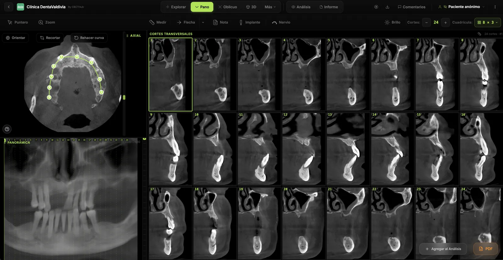
- Orthogonal MPR (axial, sagittal, coronal), then oblique planes along the long axis of the root (or the suspected canal).
- Hard-tissue window/level—do not judge canals on a soft-tissue preset. If you work in a CBCT viewer, master the basics of the viewer and measurements and notes.
- Walk the canal in thin slices looking for bifurcations, isthmuses, transportation, and incomplete fills.
- Periodontium and cortices: dehiscences, fenestrations, bone patterns suggestive of fracture.
- Neighboring structures only within the FOV: sinus, buccal/lingual cortex; IAN when included and surgery requires it.
- Millimeter measurements when they change the decision (lesion length, distance to cortex or mandibular canal).
- Correlate with clinical findings and 2D. CBCT does not automatically outrank percussion or probing.
- Document uncertainty. “No fracture line seen; metal artifact limits…” beats a forced yes/no.
To share the volume with a colleague without fighting the vendor CD, an online CBCT viewer workflow reduces friction (open without installing); that is reading logistics, not a substitute for a report.
Training, responsibility, and AI (no product promises)
The 2025 Update emphasizes training. Whoever orders or acquires CBCT must ensure competent interpretation of the FOV—by a trained clinician or by referral to oral and maxillofacial radiology. AI appears as a possible support for diagnostic consistency; it does not mean a given viewer automatically detects missed canals, fractures, or resorption. If AI exists in your environment, keep it subordinate to clinical correlation.
What this article is not
It does not list all 12 recommendations one by one (see the full JOE/OOOO text). It invents neither µSv doses nor sensitivity/specificity percentages. It is not a software pitch or a user-guide rewrite, and it does not replace local radiation rules or clinical judgment for the individual patient.
Frequently asked questions
Should CBCT be ordered for every endodontic retreatment?
No. The 2025 AAE/AAOMR framework reiterates selective use. It is justified when clinical findings and 2D imaging cannot locate the cause of failure (unresolved anatomy, resorption, suspected fracture, surgical planning). If a periapical already supports a clear, safe plan, CBCT is not mandatory “chart completion.”
What FOV should I request for a molar with a suspected missed canal?
Usually a limited FOV centered on that tooth (or the smallest field that answers the question), with resolution suited to canal detail. Avoid a both-jaws FOV “in case something else appears”—that conflicts with ALADAIP.
Does a negative CBCT rule out a vertical root fracture?
Not absolutely. Metal artifact and effective slice thickness can hide fine lines; bone pattern and clinical signs (narrow probing defect, sinus tract, failure pattern) often matter as much as “seeing the crack.” Document limitations.
Are lead aprons and thyroid collars still required for CBCT?
The 2025 statement aligns with recent ADA/AAOMR findings that they are no longer generally recommended for CBCT, but local requirements may still apply. Follow your jurisdiction and center policy.
Does artificial intelligence replace the interpreter in endodontics?
No. The 2025 Update mentions AI as a possible support for interpretation. Clinical responsibility and correlation with the examination remain with the professional.
References
- Sousa Melo SL, Fayad MI, Gohel A, et al. AAE and AAOMR Joint Position Statement: Use of Cone-Beam Computed Tomography in Endodontics 2025 Update. J Endod. 2026;52(1):4-13. doi: 10.1016/j.joen.2025.09.008. Also published as Oral Surg Oral Med Oral Pathol Oral Radiol. 2026;141(1):126-135. doi: 10.1016/j.oooo.2025.09.013. PubMed PMID: 41407481.
- AAE and AAOMR Joint Position Statement: Use of Cone Beam Computed Tomography in Endodontics 2015 Update. J Endod. 2015;41(9):1393-1396. doi: 10.1016/j.joen.2015.07.013. (Parallel publication: Oral Surg Oral Med Oral Pathol Oral Radiol. 2015;120(4):508-512. doi: 10.1016/j.oooo.2015.07.033.)
- European Commission. Radiation Protection No 172: Cone beam CT for dental and maxillofacial radiology. Evidence-based guidelines (SEDENTEXCT). 2012. PDF.
- Patel S, Durack C, Abella F, et al. European Society of Endodontology position statement: The use of CBCT in Endodontics. Int Endod J. 2014;47(6):502-504. doi: 10.1111/iej.12267.
- American Association of Endodontists. AAE and AAOMR Release Joint Position Statement on the Use of CBCT in Endodontics (press release). 14 Jan 2026. newsroom.aae.org.
Try CBCTHub for free
Upload, view, and share DICOM scans in the cloud. Nothing to install.
Create free accountRelated articles
Dental CBCT in the cloud: day-to-day storage and sharing in clinic
What “CBCT in the cloud” means in clinic: store the original DICOM, access it from multiple chairs, and share with access control — without mistaking it for a full PACS or a WhatsApp ZIP.
Browser DICOM viewer vs desktop: what works for CBCT in clinic
Open CBCT in the browser or install a desktop viewer? A clinical decision matrix: MPR performance, offline use, Mac/Windows mix, sharing without CDs, and when native software still wins.
Dental DICOM software: a checklist for choosing a CBCT viewer
Before adopting a dental DICOM or CBCT viewer in the clinic: checklist for interoperability, MPR, mm measurements, share without CD, privacy, and TCO.