CBCT vs Panoramic Radiograph: When to Use Each

· Updated

Understanding the Imaging Modalities
Panoramic radiographs and cone beam computed tomography represent two of the most commonly used imaging modalities in dentistry. Understanding their respective strengths, limitations, and clinical applications enables clinicians to make appropriate imaging selections that optimize diagnosis while minimizing radiation exposure.
Panoramic Radiographs: Traditional and Valuable
Panoramic imaging has been a cornerstone of dental diagnosis for decades. A panoramic radiograph provides a single-plane, low-resolution view of the entire dentition and supporting structures.
Advantages of Panoramic Imaging:
- Lower radiation dose compared to CBCT
- Quick acquisition and easy patient positioning
- Excellent for general screening and survey
- Ideal for detecting widespread pathology
- Cost-effective and widely accessible
- Good visualization of dental eruption and development
Limitations:
- Flat 2D representation of 3D anatomy
- Magnification and distortion of structures
- Poor visualization of small lesions
- Limited soft tissue contrast
- Superimposition of anatomical structures complicates interpretation
CBCT: Three-Dimensional Precision
CBCT vs panoramic differences are substantial when three-dimensional assessment becomes clinically necessary. CBCT provides isotropic volumetric data enabling multiplanar reconstruction.
Advantages of CBCT:
- True three-dimensional visualization without superimposition
- Precise measurement of bone dimensions and anatomy
- Superior lesion detection and characterization
- Detailed assessment of tooth position and root morphology
- Excellent for surgical planning
- Reduced magnification and distortion
Limitations:
- Higher radiation dose than panoramic radiographs
- Longer acquisition time
- Increased equipment and software costs
- Requires specialized viewer software
- Larger file sizes
Clinical Decision-Making Framework
Choose panoramic radiography when:
- Screening for dental disease and development
- Assessing overall jaw and tooth relationships
- Routine follow-up of known conditions
- Severe radiation concerns (pediatric patients, pregnancy)
- Initial evaluation in asymptomatic patients
Select CBCT when:
- Planning implant placement
- Evaluating impacted teeth or complex extractions
- Assessing TMJ disorders
- Diagnosing complex pathology
- Presurgical planning for orthognathic or oral surgery procedures
- Orthodontic assessment requiring precise skeletal measurements
- Evaluating bone graft sites
- Investigating persistent symptoms with negative panoramic findings
Radiation Dose Considerations
While CBCT doses vary by field of view and acquisition protocol, a full-mouth CBCT typically delivers 50-100 microsieverts, compared to 2-5 microsieverts for panoramic radiography. Risk-benefit analysis should guide selection, particularly in pediatric patients where dose reduction is paramount.
The Complementary Approach
Dental radiography best practices often employ both modalities. Initial screening may use panoramic imaging, with CBCT reserved for cases requiring detailed three-dimensional assessment. This sequential approach minimizes radiation exposure while ensuring adequate diagnostic information.
Technology Integration
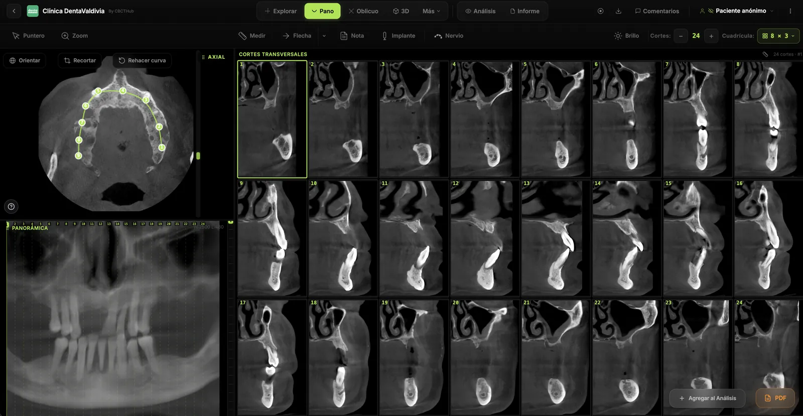
Modern imaging workflows often extract panoramic-equivalent images from CBCT data for comparison with traditional radiographs. This capability further reduces the need for separate panoramic imaging in CBCT-using practices.
Conclusion
Neither panoramic nor CBCT imaging is universally superior—each serves distinct clinical purposes. Judicious selection based on clinical questions, patient factors, and diagnostic needs ensures optimal outcomes while maintaining appropriate radiation safety.
Try CBCTHub for free
Upload, view, and share DICOM scans in the cloud. Nothing to install.
Create free accountRelated articles
Browser DICOM viewer vs desktop: what works for CBCT in clinic
Open CBCT in the browser or install a desktop viewer? A clinical decision matrix: MPR performance, offline use, Mac/Windows mix, sharing without CDs, and when native software still wins.
How to open a CBCT without installing software: a practical online-viewer guide
You got a CD, ZIP, or link with a CBCT and no manufacturer viewer. How to open the DICOM in the browser, measure in mm, and what to do when it fails.
Mandibular canal and inferior alveolar nerve on CBCT: anatomy, variants, and pre-surgical measurement
Locate the mandibular canal and IAN on CBCT: mental loop, bifid/retromolar canal, measurement checklist, and when to escalate from panoramic (ALADA).