Dental DICOM software: a checklist for choosing a CBCT viewer
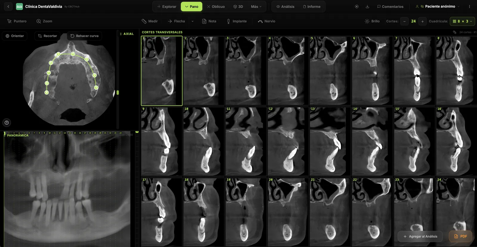
Choosing dental DICOM software is not the same as buying a full PACS or signing an infrastructure contract. For most dentists and specialists (implantology, endodontics, orthodontics, oral surgery, oral radiology), the real decision is closer to home: which CBCT / DICOM viewer you will use every day to open volumes, measure in millimeters, review the full dataset, and share studies without depending on the manufacturer’s CD.
This article is a practical decision checklist. It does not rank named competitors or invent a “winner” table. The goal is to evaluate any candidate—desktop, networked, or browser-based—with the same clinical and technical criteria before you adopt it in clinic.
Companion posts cover “what is an online viewer” and how to open a CBCT without installing the manufacturer executable. Here the focus is different: selection criteria for viewing software, not a PACS purchase guide.
1. DICOM interoperability (before the pretty UI)
DICOM (Digital Imaging and Communications in Medicine) is the NEMA standard for medical imaging. In dentistry, Farman and colleagues long argued that interoperability protects the most valuable clinical asset: patient data against proprietary software obsolescence and vendor silos.
Checklist
- [ ] The vendor publishes a DICOM Conformance Statement (which SOP Classes it imports/exports).
- [ ] You can import at least CT Image Storage and, ideally, Enhanced CT (multi-frame) when the scanner delivers it that way.
- [ ] Cone-beam export leaves the site as standard DICOM, not only a closed brand format.
- [ ] Useful metadata is preserved (patient, study, series, spacing/pixel size); the volume is not flattened to JPEG.
- [ ] You run a real test with 2–3 volumes from different brands in your referral network: paper conformance does not guarantee practical interoperability.
Ask the vendor: “Can I open the DICOM that center X sends without their viewer?” If the answer is “only if you install our package,” you are evaluating a silo, not an interoperable viewer.
2. Multi-vendor CBCT import
In real clinics, Carestream, Planmeca, Vatech, Sirona, Morita, NewTom and others all arrive. A useful viewer does not require every referrer to share one workstation brand.
Checklist
- [ ] Imports DICOM folders / ZIP / DICOMDIR without hidden proprietary conversion steps.
- [ ] Finds the correct volumetric series when scouts or 2D reconstructions sit in the same study.
- [ ] Does not force you to burn a CD with the manufacturer installer just to “see” the case.
- [ ] Documents known limitations (transfer syntax, Enhanced CT, incomplete series) in plain language for the team.
If your workflow depends on external referrals, this item outweighs any demo built on a single “perfect” manufacturer dataset.
3. MPR, cross-sections, and clinical navigation
EADMFR / SEDENTEXCT guidance and the AAOMR executive opinion stress that CBCT is not “four PDF snapshots”: the entire dataset must be reviewable. A viewer that only shows a fixed reconstruction fails basic clinical use.
Checklist
- [ ] Synchronized MPR: navigable axial, sagittal, and coronal planes.
- [ ] Oblique / cross-sectional views useful for implants and nerve pathways (by specialty).
- [ ] Panoramic or curved reconstruction if you use it in planning (not mandatory for everyone, but reliable if offered).
- [ ] Clear zoom, window/level, and anatomic orientation (avoid L/R confusion).
- [ ] Smooth scrolling through hundreds of slices—not only a fast first image.
For surgery and implantology, prioritize stable cross-sections and reproducible measurements. For endodontics, perceived spatial resolution and fine scroll. For orthodontics, large FOV and coherent cephalic orientation when relevant.
4. Measurement tools you actually use chairside
A demo packed with “AI” does not replace a trustworthy millimeter ruler.
Checklist
- [ ] Distance in mm calibrated from DICOM metadata (coherent Pixel Spacing / Slice Thickness).
- [ ] Angles and, if offered, area or relative density with the caveat that CBCT is not absolute densitometry like a fully Hounsfield-calibrated medical CT on every unit.
- [ ] Exportable annotations or captures with visible scale for the chart / report.
- [ ] A documented way to verify a known measurement for each new scanner type you receive.
Before trusting an implant plan, measure a plausible anatomic distance and compare with the radiologist’s report or another reference workstation.
5. Sharing and export without a CD
Manufacturer CDs still exist, but modern flow expects a DICOM ZIP, a secure link, or center integration. Farman warned that proprietary formats trap data; open export frees it.
Checklist
- [ ] Full DICOM export (not screenshots only).
- [ ] Sharing via an access-controlled link (expiry, PIN, or authentication)—not only a 500 MB email attachment.
- [ ] Capture / visual report export for the patient or referrer without replacing the volume.
- [ ] No dependence on a single PC that still has the manufacturer installer to “open what we sent.”
Sharing is not long-term archiving: multi-year legal retention needs a retention policy plus backup; the viewer should fit that policy, not invent it ad hoc.
6. Privacy and basic compliance
Dental images are health data. In the U.S., HIPAA’s minimum-necessary and reasonable-safeguards framing applies; in the EU and many LATAM contexts, GDPR and local laws apply to transfers. This is not legal advice—demand transparency from the vendor.
Checklist
- [ ] Encryption in transit (HTTPS/TLS) for any network/cloud upload or viewing.
- [ ] Control over who opens the study (account, scoped link, access logging if applicable).
- [ ] Clear policy on where data are stored and for how long.
- [ ] Ability to delete / expire shared links.
- [ ] Contract or DPA when the vendor processes personal data on your behalf.
- [ ] A workflow that does not push PHI through WhatsApp or personal Drive without controls.
A “free public cloud viewer with no account” may be fine for an anonymized teaching case; it is not automatically suitable for production clinical identifiers.
7. Install on every PC vs. open in the browser
A deeper browser-versus-desktop analysis belongs to a later cluster piece. Here, only what affects adoption:
Checklist
- [ ] How many seats need to see CBCT (operatory, planning box, specialist at home)?
- [ ] Mixed Mac and Windows? Tablets?
- [ ] Can you update software without visiting every PC?
- [ ] Is licensing per seat, per clinic, or per use?
- [ ] If browser-based: partial offline mode or stable network required?
- [ ] If desktop: who maintains drivers, GPU, and restores after a wipe?
An excellent viewer that only runs on one Windows box recreates the same bottleneck as the CD.
8. Performance with large volumes
A large-FOV CBCT can exceed hundreds of MB. Delay is not just annoyance—it interrupts the visit.
Checklist
- [ ] Load time measured on your typical files (not an 80 MB demo).
- [ ] Behavior on clinic Wi‑Fi versus wired network.
- [ ] Observable CPU/RAM/GPU use (does the rest of the PC freeze?).
- [ ] Strategy for incomplete series or truncated ZIPs (clear message, not a blank screen).
- [ ] Transfer/compression that does not destroy spatial calibration.
Test the real worst case: Friday 6 p.m., patient in the chair, 450 MB ZIP over reception Wi‑Fi.
9. Support, training, and clinical responsibility
The AAOMR reminds practitioners that whoever obtains the CBCT is responsible for interpreting the volume (or referring to oral radiology). Software does not remove that duty; it can make full-volume review easier or harder.
Checklist
- [ ] Team onboarding in <1–2 sessions for staff who open studies daily.
- [ ] Support in your language and reasonable hours.
- [ ] Updated training materials (without replacing clinical judgment).
- [ ] Clear path when a file “won’t open”: re-export DICOM, transfer syntax, correct series.
- [ ] Vendor does not market automatic diagnosis as a substitute for human review of the dataset.
SEDENTEXCT / RP 172 and EADMFR principles also stress ongoing training when new techniques or equipment are adopted—include reading software in that plan.
10. Total cost of ownership (TCO), not just the license
Checklist
- [ ] Initial license + renewals + extra seats.
- [ ] IT time / “the person who knows how to install” in the clinic.
- [ ] Cost of failing to open a referral (rework, unjustified rescan, lost appointment).
- [ ] Storage, backups, and data exit if you change vendors (lock-in).
- [ ] Minimum hardware (GPU, monitors, bandwidth).
“Cheap” software that forces reinstalls on three PCs every update—or rescans because files won’t open—gets expensive fast.
One-page checklist
- Conformance Statement + real multi-vendor test.
- Navigable MPR + verifiable mm measurements.
- DICOM export/share without a proprietary CD.
- Privacy: encryption, controlled access, clear retention.
- Deployment model that matches your seats (PC / browser).
- Performance on your large volumes.
- Support and team training.
- 2–3 year TCO, including data exit.
What this checklist is not
- Not a dental PACS buyer’s guide (servers, full HL7, hospital worklist).
- Not a substitute for ALARA justification or CBCT indication (EADMFR / SEDENTEXCT).
- Not a brand comparison or “best free viewers” roundup.
- Not an inventory of one product’s features—evaluate with evidence in your workflow.
References
- Horner K, Islam M, Flygare L, Tsiklakis K, Whaites E. Basic principles for use of dental cone beam computed tomography: consensus guidelines of the European Academy of Dental and Maxillofacial Radiology. Dentomaxillofac Radiol. 2009;38(4):187-195. doi:10.1259/dmfr/74941012
- European Commission. Radiation Protection No 172. Cone Beam CT for Dental and Maxillofacial Radiology: Evidence-Based Guidelines (SEDENTEXCT). Luxembourg: Publications Office of the EU; 2012. PDF
- Carter L, Farman AG, Geist J, et al. American Academy of Oral and Maxillofacial Radiology executive opinion statement on performing and interpreting diagnostic cone beam computed tomography. Oral Surg Oral Med Oral Pathol Oral Radiol Endod. 2008;106(4):561-562. doi:10.1016/j.tripleo.2008.07.007
- Farman AG. Raising standards: Digital interoperability and DICOM. Oral Surg Oral Med Oral Pathol Oral Radiol Endod. 2005;99(5):525-526. doi:10.1016/j.tripleo.2005.03.001
- Farman AG. Applying DICOM to dentistry. J Digit Imaging. 2005;18(1):23-27. doi:10.1007/s10278-004-1029-z
- Farman AG, Lapp RP. Image file interoperability for data protection, communication and trans-system connectivity. Orthod Craniofac Res. 2003;6(Suppl 1):151-155. doi:10.1034/j.1600-0544.2003.248.x
- National Electrical Manufacturers Association (NEMA). DICOM Standard (current edition). Available at: https://dicom.nema.org/medical/dicom/current/
- U.S. Department of Health & Human Services. Minimum Necessary Requirement (HIPAA Privacy Rule). https://www.hhs.gov/hipaa/for-professionals/privacy/guidance/minimum-necessary-requirement/index.html
Try CBCTHub for free
Upload, view, and share DICOM scans in the cloud. Nothing to install.
Create free accountRelated articles
Online CBCT viewer: what it is and when to use it in clinic
What an online CBCT viewer is, when to use it vs desktop or CD, DICOM tools (MPR, measurements, share link) and basic privacy in clinic.
How to open a CBCT without installing software: a practical online-viewer guide
You got a CD, ZIP, or link with a CBCT and no manufacturer viewer. How to open the DICOM in the browser, measure in mm, and what to do when it fails.
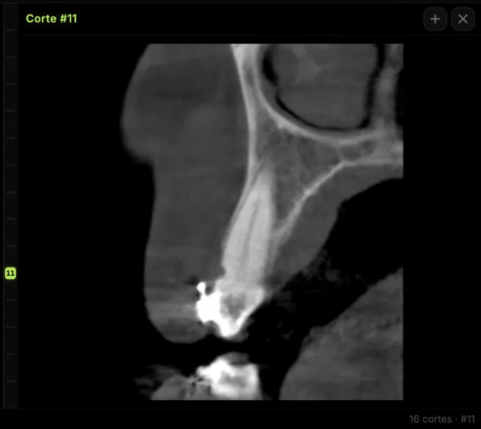
Mandibular canal and inferior alveolar nerve on CBCT: anatomy, variants, and pre-surgical measurement
Locate the mandibular canal and IAN on CBCT: mental loop, bifid/retromolar canal, measurement checklist, and when to escalate from panoramic (ALADA).