What Is CBCT and Why Every Dental Practice Needs It


Understanding CBCT Technology
Cone Beam Computed Tomography (CBCT) has transformed the landscape of dental imaging. Unlike traditional 2D radiographs, CBCT provides three-dimensional visualization of dental and maxillofacial structures, enabling clinicians to make more informed diagnostic and treatment decisions.
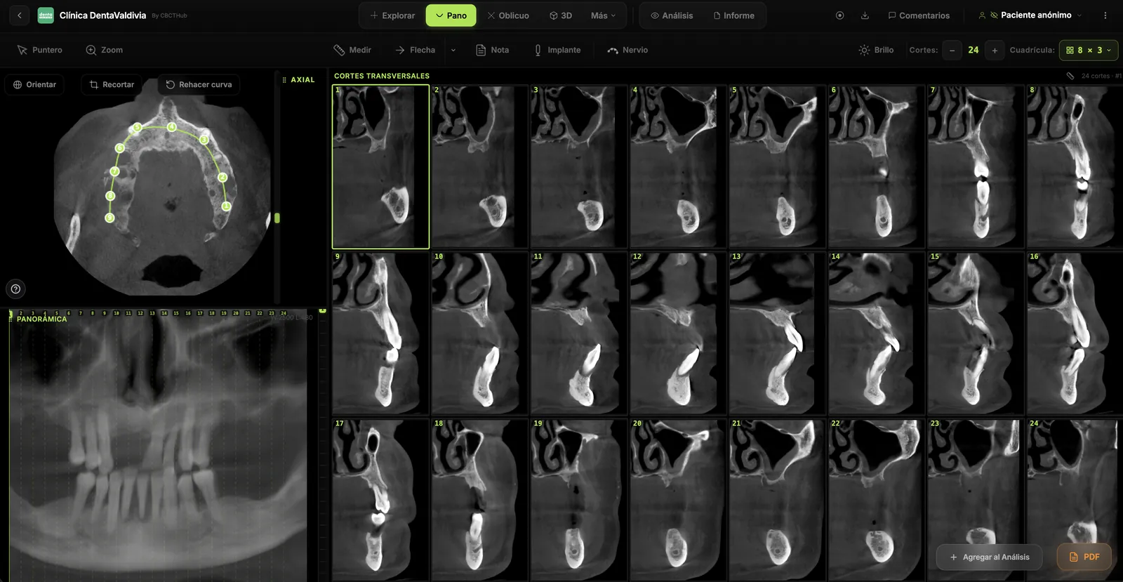
CBCT dental imaging uses a cone-shaped X-ray beam to capture data from a 360-degree rotation around the patient's head. This data is then reconstructed into cross-sectional images—axial, sagittal, and coronal views—and full 3D volumetric models.
Key Advantages of CBCT in Modern Dentistry
The benefits of implementing CBCT in your practice are substantial:
- Enhanced Diagnostic Accuracy: 3D imaging reveals anatomical details impossible to see in 2D radiographs, improving diagnosis of bone pathology, impacted teeth, and TMJ disorders.
- Improved Treatment Planning: Whether for implant placement, orthodontics, or surgical extractions, CBCT provides precise measurements and spatial relationships.
- Reduced Treatment Time: Better visualization leads to more efficient procedures and fewer complications.
- Patient Communication: 3D images help patients understand complex conditions and treatment options, increasing case acceptance.
- Legal Protection: Comprehensive imaging documentation protects practitioners in liability situations.
Clinical Applications Across Specialties
CBCT is invaluable across multiple dental disciplines. General dentists use it for implant assessment, wisdom tooth evaluation, and detecting hidden caries. Orthodontists rely on CBCT for precise skeletal measurements and airway assessment. Oral surgeons depend on it for complex extractions and bone grafting procedures.
Periodontists utilize CBCT to evaluate bone loss and plan regenerative procedures. Prosthodontists benefit from accurate bone anatomy when planning implant restorations. Endodontists use CBCT to identify calcified canals and complex anatomies.
Integration with Digital Workflows
Modern CBCT systems integrate seamlessly with digital practice management and imaging software. CBCTHub and similar online DICOM viewers enable practitioners to access and share CBCT data across multiple devices and locations. This cloud-based approach eliminates the need for CD-based distribution and enables real-time collaboration among clinical teams.
Conclusion
Implementing CBCT in your dental practice represents a significant investment in diagnostic capability and patient care quality. As digital dentistry continues to evolve, cone beam imaging has become less of a luxury and more of a necessity for competitive, evidence-based practice. Whether you're just starting with CBCT or optimizing existing imaging workflows, the three-dimensional perspective this technology provides is invaluable.
Try CBCTHub for free
Upload, view, and share DICOM scans in the cloud. Nothing to install.
Create free accountRelated articles
Browser DICOM viewer vs desktop: what works for CBCT in clinic
Open CBCT in the browser or install a desktop viewer? A clinical decision matrix: MPR performance, offline use, Mac/Windows mix, sharing without CDs, and when native software still wins.
How to open a CBCT without installing software: a practical online-viewer guide
You got a CD, ZIP, or link with a CBCT and no manufacturer viewer. How to open the DICOM in the browser, measure in mm, and what to do when it fails.
Mandibular canal and inferior alveolar nerve on CBCT: anatomy, variants, and pre-surgical measurement
Locate the mandibular canal and IAN on CBCT: mental loop, bifid/retromolar canal, measurement checklist, and when to escalate from panoramic (ALADA).