Endodontic CBCT indications: a guide to complex cases

Endodontic CBCT indications are limited to cases in which clinical examination and periapical radiographs fail to answer the diagnostic question and three-dimensional information could change treatment: contradictory or nonspecific signs and symptoms, complex anatomy, calcified canals, nonhealing of previous treatment, apical surgery planning, suspected vertical root fracture, resorption and dentoalveolar trauma[1]. CBCT is used selectively, never routinely, with a limited field of view (FOV) and under the ALADAIP principle[2].
Key points
- Intraoral radiography remains the imaging modality of choice for the endodontic patient; limited FOV CBCT is reserved for questions 2D imaging cannot answer[1].
- The 2025 update of the AAE/AAOMR joint position statement condenses the recommendations from 14 to 12 and confirms that CBCT should be used selectively, not routinely, under ALADAIP[2].
- In patients referred under European criteria, CBCT changed the treatment plan in 53% of cases[3]; its influence is significant mainly in high-difficulty cases[4].
- In a study of 1,397 limited FOV volumes, 23.04% of treated premolars and molars had a missed canal, and those teeth were 4.38 times more likely to show a periapical lesion[5].
- For vertical root fracture in root-filled teeth, pooled CBCT sensitivity and specificity were only 0.752 and 0.652[6].
- The three-dimensional classification of external cervical resorption describes height, circumferential spread and proximity to the canal, and relies on CBCT assessment[7].
When is CBCT indicated in endodontics?
The right question is not "is this case complex?" but "what information am I missing, and can a 3D scan provide it?". The joint position statement of the American Association of Endodontists (AAE) and the American Academy of Oral and Maxillofacial Radiology (AAOMR) starts from a clear premise: intraoral radiographs are the modality of choice for evaluating the endodontic patient, and limited FOV CBCT is considered when clinical findings and 2D imaging are contradictory or inconclusive[1]. The 2025 update keeps that logic, stresses training in CBCT use and interpretation, and applies ALADAIP (As Low As Diagnostically Acceptable, being Indication-oriented and Patient-specific)[2].
The European Society of Endodontology (ESE) published its own update in 2019, noting that since 2014 more clinical studies have confirmed the positive impact of CBCT on treatment planning and decision-making when reviewing cases[8]. Both guidelines agree on the starting point: clinical examination and two periapicals at different angles first; the volume only if the question remains open.
Endodontic CBCT indications according to AAE/AAOMR
The 2015 statement listed 14 recommendations that remain the working reference for defining complex cases[1]; the 2025 version consolidates them into 12 while keeping selective use[2]. The table summarizes, by scenario, which question the volume answers and what the radiologist should report.
| Clinical scenario | Question CBCT answers | What to report |
|---|---|---|
| Contradictory or nonspecific signs and symptoms[1] | Is there a periapical lesion or nonendodontic pathology the periapical misses? | Tooth and root involved, extent, cortical involvement, relation to sinus or mandibular canal |
| Complex anatomy: mandibular incisors, premolars, molars, dental anomalies[1] | How many canals are there and where do they split? | Number of roots and canals, level of division, curvatures, dens invaginatus or other anomalies |
| Calcified canals[1] | Where is the residual canal lumen? | Level at which the canal becomes visible and its position relative to coronal landmarks |
| Nonhealing and retreatment[1] | Why did it fail? Nonsurgical, surgical or extraction? | Missed canals, overextension, separated instruments, perforations, lesion extent |
| Apical surgery planning[1] | Where are the apices and what lies nearby? | Distances to buccal cortex, mandibular canal, mental foramen and maxillary sinus |
| Suspected vertical root fracture with inconclusive clinical and 2D findings[1] | Is there a fracture line or a compatible bone pattern? | Direct or indirect finding, with artifact limitations stated explicitly |
| Internal and external resorption[1] | Internal or external? Is it treatable? | Location, vertical and circumferential extent, communication with the canal |
| Localized dentoalveolar trauma[1] | Root fracture, luxation or alveolar fracture? | Fracture level and orientation, displacement, integrity of the cortical plates |
For a commented reading of the guideline and its recent changes, see Endodontic CBCT: the AAE/AAOMR 2025 statement, and for case examples of how field size matters, when a small FOV changes the outcome.
How much does CBCT change endodontic decision-making?
Justifying a scan depends on therapeutic efficacy, not just diagnostic accuracy. In a prospective study by Mota de Almeida et al., only 4% of patients referred to two endodontic clinics were examined with CBCT following European criteria; in that group, the most frequent reason was to differentiate pathology from normal anatomy (45% of cases), and the treatment plan changed in 53% of patients[3].
Rodríguez et al. presented 30 cases of varying difficulty to prosthodontists, endodontists, oral surgeons and periodontists, first without and then with a small-volume CBCT. The scan significantly influenced the treatment plan only in high-difficulty cases, and after viewing it the extraction option increased[4]. The practical message is twofold: CBCT pays off where complexity is real, and additional information can also push toward more radical decisions, so the report must be precise and avoid overstating equivocal findings.
| Study | Design | Main finding |
|---|---|---|
| Patel et al., 2009[9] | Artificial lesions in dry human jaws | Sensitivity 24.8% with digital periapical vs 100% with CBCT |
| Mota de Almeida et al., 2014[3] | Prospective, before/after CBCT | Plan changed in 53% of patients |
| Karabucak et al., 2016[5] | Retrospective, 1,397 volumes | 23.04% missed canals; OR 4.38 for lesion |
| Talwar et al., 2016[6] | Systematic review and meta-analysis | Low accuracy for vertical fracture in root-filled teeth |
| Rodríguez et al., 2017[4] | 30 cases assessed by specialists | Significant impact only in high-difficulty cases |
The Patel et al. study is ex vivo with artificial defects, but it illustrates the mechanism: anatomical noise and projection geometry hide lesions in 2D, while CBCT lets the clinician select the most relevant plane[9]. In patients, this advantage translates into indications such as persistent pain with an unremarkable periapical.
Retreatment planning: missed canals, perforations and separated instruments
Nonhealing of previous treatment is one of the highest-yield indications. Karabucak et al. reviewed 1,397 limited FOV volumes (76 µm or 125 µm voxels) and found missed canals in 23.04% of treated premolars and molars; incidence was highest in maxillary molars (40.1%) and lowest in maxillary premolars (9.5%), reaching 46.5% in tooth #14 and 41.3% in tooth #3 (Universal numbering)[5]. Teeth with a missed canal were 4.38 times more likely to be associated with a lesion, and the authors recommend examining a limited FOV CBCT before any retreatment[5].
For nonsurgical retreatment, AAE/AAOMR recommends limited FOV CBCT to assess complications such as overextended obturation material, separated instruments and perforation localization[1]. When reading:
- Scroll axial slices from the pulp chamber to the apex looking for a canal lumen without radiopaque material, especially in the mesiobuccal root of maxillary molars.
- Align an oblique slice with each root axis to follow the full canal and avoid mistaking an apical split for an independent canal.
- For a suspected perforation, describe its level (cervical, middle or apical), the root surface involved and any adjacent bone involvement.
- Distinguish overfilling from material extruded into the sinus or mandibular canal, and measure the distance.
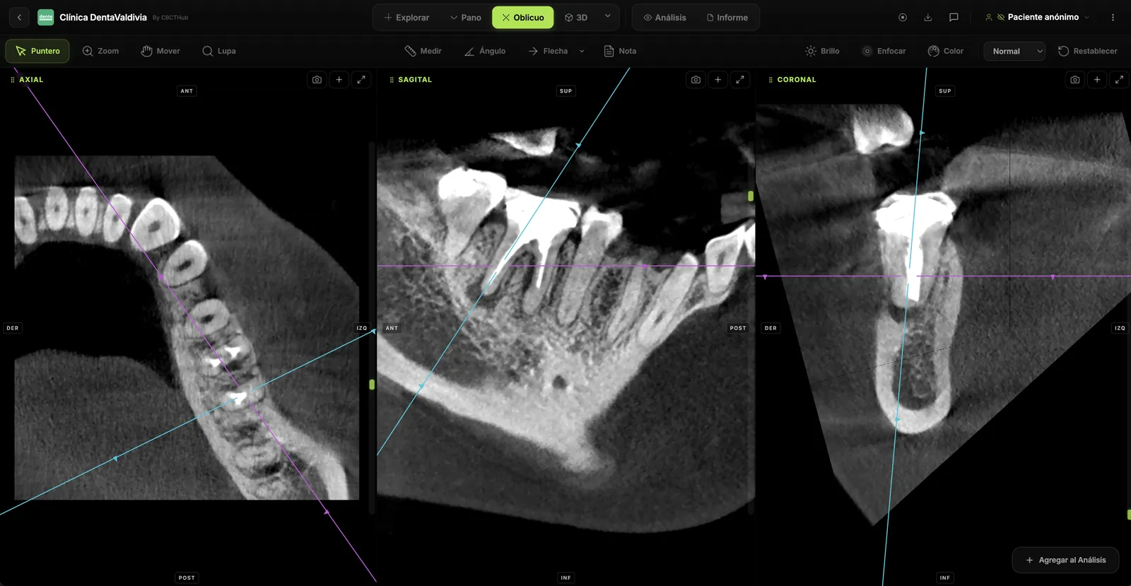
Clinical pearl: gutta-percha and metal posts cause beam hardening and dark streaks that mimic canals or fracture lines. Before reporting a missed canal, confirm it in at least two planes and across several contiguous slices.
Vertical root fracture on CBCT: realistic expectations
Suspected vertical root fracture is an accepted indication when clinical and 2D findings are inconclusive[1], but expectations need calibrating. In the Talwar et al. meta-analysis, pooled CBCT sensitivity and specificity were 0.776 and 0.946 in unfilled teeth, dropping to 0.752 and 0.652 in root-filled teeth[6]. Better accuracy in unfilled teeth was observed particularly with a 0.2 mm voxel[6]. Artifact recognition is covered in depth in our CBCT artifacts field guide.
A specificity of 0.652 in root-filled teeth means a relevant share of reported "fractures" do not exist. The report should therefore separate a fracture line visible in several planes from findings that are merely compatible, and explicitly state artifacts from obturation material or posts. A false positive here can lead to an unnecessary extraction.
Resorption: localization and three-dimensional classification
Limited FOV CBCT is the modality of choice for localizing and differentiating internal and external resorption and for determining treatment and prognosis[1]. For external cervical resorption, the three-dimensional classification by Patel et al. structures the description along three axes[7]:
| Axis | Categories |
|---|---|
| Height | 1: at CEJ level or coronal to the bone crest; 2: coronal third, apical to the crest; 3: middle third; 4: apical third |
| Circumferential spread | A: ≤90°; B: ≤180°; C: ≤270°; D: >270° |
| Proximity to the canal | d: confined to dentine; p: probable pulpal involvement |
Reporting in this format (for example, "2Bd") lets the endodontist and surgeon speak the same language when choosing an external, internal or extraction approach[7].
Dentoalveolar trauma and apical surgery planning
In localized dentoalveolar trauma, limited FOV CBCT is indicated for diagnosing and managing root fractures, luxation, displacement and localized alveolar fractures, provided there are no other maxillofacial injuries requiring another modality[1]; see also CBCT in dental trauma. For apical surgery, the volume localizes the apices and shows their proximity to adjacent anatomical structures[1]: measure buccal cortical thickness, apex-to-mandibular canal or mental foramen distance, and the relation to the maxillary sinus floor.
When should you NOT order CBCT in endodontics?
- As a routine exam for every endodontic patient: the 2025 update expressly rules this out[2].
- For immediate postoperative imaging, where intraoral radiography is the modality of choice[1].
- To assess healing in the absence of signs or symptoms, unless the initial diagnosis was made with CBCT[1].
- When no possible answer from the volume would change the treatment plan.
The 2025 statement aligns with the ADA and AAOMR in that lead aprons and thyroid collars are no longer recommended for CBCT, although local regulations may still require them[2].
Acquisition and reading protocol: what the report should include
AAE/AAOMR recommends an FOV only slightly larger than the region of interest, the smallest voxel that answers the question, the lowest mA appropriate for patient size, and low-dose protocols when the question does not require high resolution; larger FOVs are reserved for extensive, multifocal or possibly nonendodontic lesions[1]. Volume quality drives performance: check noise, motion and artifacts with the CBCT image quality checklist.
A useful report for a complex endodontic case should include:
- The referrer's clinical question and the protocol (FOV, voxel, unit).
- The main finding per tooth and root, with measurements in mm where relevant.
- Relation to neighboring structures: maxillary sinus, mandibular canal, mental foramen, cortical plates.
- Limitations: artifacts, motion, insufficient resolution for the question.
- Findings outside the region of interest within the acquired volume.
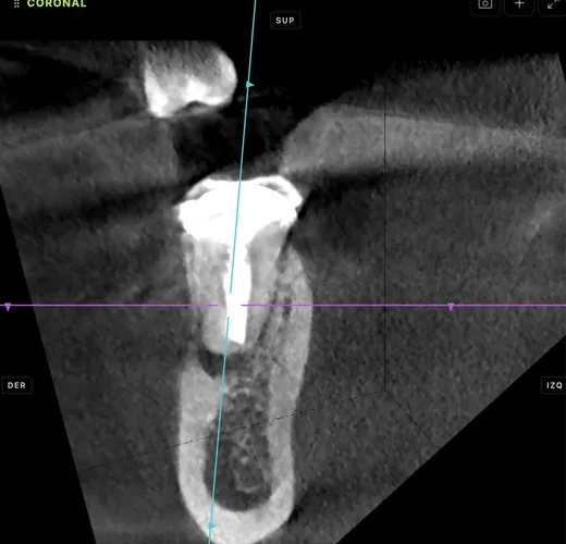
How to do it in CBCTHub
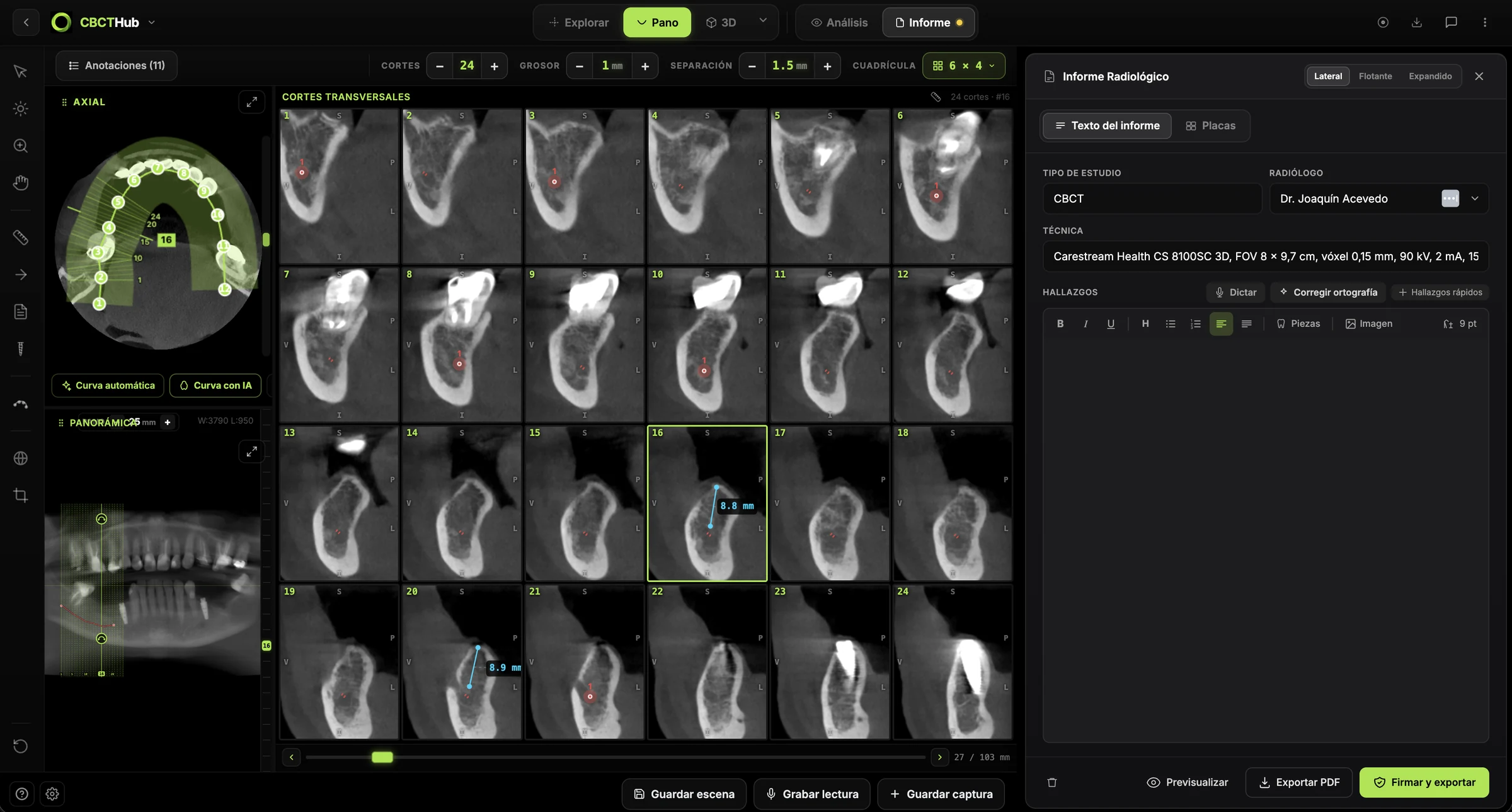
CBCTHub lets you read, measure, report and deliver an endodontic CBCT in the browser, with DICOM processed locally:
- Open the study from the link sent by the imaging center, or load the DICOM in the viewer (compatible with Carestream, Planmeca, Vatech, Sirona, Morita, NewTom and others).
- Use the endodontic module to align oblique slices with each root axis and follow the entire canal.
- Measure lesion extent and its distance to the sinus, mandibular canal or cortex in mm.
- Leave comments linked to the exact slice showing the finding for the referring endodontist.
- Write the report in the editor next to the viewer and export image sheets as PDF.
Radiologists and imaging centers can deliver the study and report to referrers by link, without CDs, on a study page carrying the center's branding. Endodontists and dentists can open and measure the CBCT sent by the center directly in the browser. Try the CBCT viewer for free without an account or create an account at CBCTHub.
Conclusion
Limited FOV CBCT adds the most in endodontics when the case is truly complex: retreatment, missed canals, resorption, apical surgery, trauma and symptoms unexplained by 2D imaging. Its clearest limitation is vertical fracture in root-filled teeth. The indication should start from a specific question, and the report should answer it with measurements, anatomical context and stated limitations. This content is intended for professionals and does not replace clinical judgment.
Frequently asked questions
Does CBCT replace periapical radiography in endodontics?
No. According to the AAE/AAOMR joint position statement, intraoral radiography remains the modality of choice for evaluating the endodontic patient and for immediate postoperative imaging. Limited FOV CBCT is added when clinical findings and 2D imaging are contradictory or inconclusive and three-dimensional information could change treatment. The 2025 update stresses that CBCT should not be used routinely.
Which FOV and voxel size suit an endodontic CBCT?
A limited FOV only slightly larger than the region of interest, with the smallest voxel that answers the clinical question and the lowest mA appropriate for the patient. Larger fields are reserved for extensive, multifocal or possibly nonendodontic lesions. For vertical fracture in unfilled teeth, the best performance in the Talwar meta-analysis was observed with a 0.2 mm voxel.
Can CBCT reliably detect a vertical root fracture?
Only partially. In unfilled teeth, pooled sensitivity and specificity were 0.776 and 0.946, but in root-filled teeth they dropped to 0.752 and 0.652 because of artifacts from gutta-percha and posts. The report should separate a fracture line seen in several planes from merely compatible findings and state which artifacts are present.
Should CBCT be ordered before endodontic retreatment?
In most premolar and molar retreatments, yes. Karabucak et al. found missed canals in 23.04% of treated teeth and a 4.38-fold higher likelihood of a lesion when a canal was left untreated. AAE/AAOMR recommends limited FOV CBCT to assess missed canals, perforations, separated instruments and overextension before choosing the approach.
What should an endodontic CBCT report include?
The clinical question, the acquisition protocol, the finding per tooth and root with measurements in mm, the relation to the maxillary sinus, mandibular canal, mental foramen and cortical plates, limitations caused by artifacts or motion, and findings outside the region of interest. For external cervical resorption, use the three-dimensional classification of height, circumferential spread and canal proximity.
References
- AAE and AAOMR Joint Position Statement: Use of Cone Beam Computed Tomography in Endodontics 2015 Update. J Endod. 2015;41(9):1393-6. doi:10.1016/j.joen.2015.07.013
- Sousa Melo SL, Fayad MI, Gohel A, Johnson BR, Kalathingal S, Mahdian M, et al. AAE and AAOMR Joint Position Statement: Use of Cone-Beam Computed Tomography in Endodontics 2025 Update. J Endod. 2026;52(1):4-13. doi:10.1016/j.joen.2025.09.008
- Mota de Almeida FJ, Knutsson K, Flygare L. The effect of cone beam CT (CBCT) on therapeutic decision-making in endodontics. Dentomaxillofac Radiol. 2014;43(4):20130137. doi:10.1259/dmfr.20130137
- Rodríguez G, Abella F, Durán-Sindreu F, Patel S, Roig M. Influence of Cone-beam Computed Tomography in Clinical Decision Making among Specialists. J Endod. 2017;43(2):194-9. doi:10.1016/j.joen.2016.10.012
- Karabucak B, Bunes A, Chehoud C, Kohli MR, Setzer F. Prevalence of Apical Periodontitis in Endodontically Treated Premolars and Molars with Untreated Canal: A Cone-beam Computed Tomography Study. J Endod. 2016;42(4):538-41. doi:10.1016/j.joen.2015.12.026
- Talwar S, Utneja S, Nawal RR, Kaushik A, Srivastava D, Oberoy SS. Role of Cone-beam Computed Tomography in Diagnosis of Vertical Root Fractures: A Systematic Review and Meta-analysis. J Endod. 2016;42(1):12-24. doi:10.1016/j.joen.2015.09.012
- Patel S, Foschi F, Mannocci F, Patel K. External cervical resorption: a three-dimensional classification. Int Endod J. 2018;51(2):206-14. doi:10.1111/iej.12824
- Patel S, Brown J, Semper M, Abella F, Mannocci F. European Society of Endodontology position statement: Use of cone beam computed tomography in Endodontics. Int Endod J. 2019;52(12):1675-8. doi:10.1111/iej.13187
- Patel S, Dawood A, Mannocci F, Wilson R, Pitt Ford T. Detection of periapical bone defects in human jaws using cone beam computed tomography and intraoral radiography. Int Endod J. 2009;42(6):507-15. doi:10.1111/j.1365-2591.2008.01538.x
Try CBCTHub for free
Upload, view, and share DICOM scans in the cloud. Nothing to install.
Create free accountRelated articles

CBCT transverse analysis: an orthodontic guide
CBCT transverse analysis: how to measure basal width and molar inclination to tell skeletal deficiency apart from dentoalveolar compensation.

TMJ CBCT: what it can assess and where it falls short
TMJ CBCT: which osseous changes it shows, what it misses compared with MRI, and how to report condylar findings using DC/TMD imaging criteria.

CBCT pre-implant assessment: what to check first
CBCT pre-implant assessment, step by step: cross-sectional measurements, the 2 mm safety margin, critical structures and what the report must include.