Detecting Periapical Pathology with CBCT: What 2D Radiographs Miss

· Updated

The Limits of 2D Periapical Imaging
Periapical radiographs have been the diagnostic standard for decades. They're quick, low-dose, and widely available. But they have a fundamental limitation: they compress a 3D structure into a 2D image. This means periapical lesions confined to cancellous bone — without involvement of the buccal or lingual cortical plate — can be invisible on conventional radiographs.
Studies consistently show that periapical radiographs require 30-50% mineral loss before a lesion becomes visible. CBCT, by contrast, can detect lesions at much earlier stages because it eliminates the superimposition of overlying structures.
What Research Shows
Multiple studies have demonstrated the superiority of CBCT for periapical pathology detection:
- CBCT detects 34% more periapical lesions than periapical radiographs in endodontically treated teeth
- Lesion size measurements on CBCT are more accurate — 2D radiographs tend to underestimate lesion dimensions
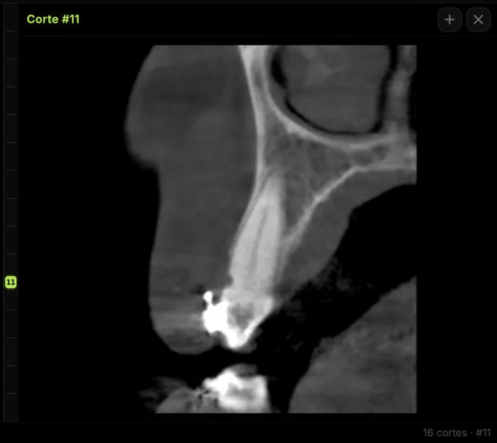
- The buccal-lingual extent of pathology, completely invisible on 2D images, is clearly shown on CBCT axial views
- CBCT can differentiate between granulomas and cysts with greater reliability, aiding treatment planning
When Does It Matter Clinically?
The additional sensitivity of CBCT for periapical pathology is most valuable in these scenarios:
Failed Root Canal Evaluation
When a patient has persistent symptoms after endodontic treatment but the periapical radiograph looks normal, CBCT often reveals a lesion in the buccal or lingual bone that was hidden on the 2D image. It can also show missed canals, separated instruments, or perforations.
Pre-surgical Assessment
Before apicoectomy, CBCT defines the exact size and extent of the lesion, its relationship to adjacent vital structures (inferior alveolar nerve, maxillary sinus, adjacent tooth roots), and the presence of any complicating factors like root fractures or extra canals.
Implant Site Evaluation
Before placing an implant at a site with a history of endodontic pathology, CBCT confirms complete resolution of any previous lesion and shows the quality and quantity of bone available for implant placement.
Unexplained Pain
When a patient presents with dental pain but no lesion is visible on conventional imaging, CBCT can reveal early pathology, cracked teeth, or atypical lesion locations that explain the symptoms.
Practical Recommendations
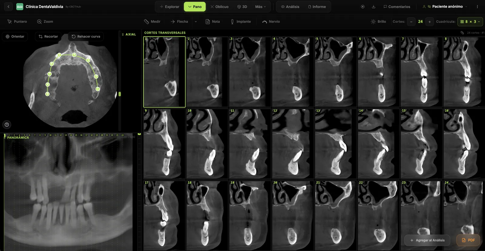
You don't need CBCT for every periapical assessment — it would be inappropriate to replace periapical radiographs as a screening tool. But when conventional images don't explain clinical findings, or when treatment decisions depend on precise lesion characterization, CBCT should be the next step. Online CBCT viewers with measurement tools and annotation capabilities make it easy to document findings and share them with the treating clinician.
Try CBCTHub for free
Upload, view, and share DICOM scans in the cloud. Nothing to install.
Create free accountRelated articles
Browser DICOM viewer vs desktop: what works for CBCT in clinic
Open CBCT in the browser or install a desktop viewer? A clinical decision matrix: MPR performance, offline use, Mac/Windows mix, sharing without CDs, and when native software still wins.
How to open a CBCT without installing software: a practical online-viewer guide
You got a CD, ZIP, or link with a CBCT and no manufacturer viewer. How to open the DICOM in the browser, measure in mm, and what to do when it fails.
Mandibular canal and inferior alveolar nerve on CBCT: anatomy, variants, and pre-surgical measurement
Locate the mandibular canal and IAN on CBCT: mental loop, bifid/retromolar canal, measurement checklist, and when to escalate from panoramic (ALADA).