CBCT for Endodontics: How 3D Imaging Improves Root Canal Diagnosis

· Updated

Why Endodontists Need CBCT
Endodontic treatment relies heavily on accurate diagnosis of root canal anatomy and periapical pathology. Traditional periapical radiographs provide a 2D snapshot, but they compress three-dimensional structures into a flat image — leading to superimposition, geometric distortion, and missed findings.
CBCT for endodontics eliminates these limitations by providing cross-sectional views at sub-millimeter resolution, allowing endodontists to see every canal, every root, and every periapical lesion in three dimensions.
Clinical Applications in Endodontics
CBCT imaging has become indispensable for several endodontic scenarios:
1. Detecting Missed Canals
One of the most common causes of endodontic failure is untreated canals. CBCT axial views clearly reveal the number and location of root canals. The second mesiobuccal canal (MB2) in upper molars, for example, is detected in over 90% of cases with CBCT compared to only 30-40% with 2D radiographs.
2. Diagnosing Vertical Root Fractures
Vertical root fractures (VRFs) are notoriously difficult to diagnose on periapical films. CBCT can reveal the fracture line, adjacent bone loss patterns, and help distinguish VRFs from other causes of localized bone loss. Sagittal and axial slices are most useful for this assessment.
3. Periapical Pathology Assessment
CBCT detects periapical lesions earlier and more accurately than 2D radiographs. Studies show that up to 35% of periapical lesions visible on CBCT are not detectable on conventional radiographs, particularly when the lesion is confined to cancellous bone without cortical plate involvement.
4. Root Resorption Evaluation
Both internal and external root resorption can be precisely mapped with CBCT. The exact location, extent, and proximity to the root canal space are clearly visible, enabling better treatment decisions about whether to treat or extract.
When to Order a CBCT for Endodontic Cases
The American Association of Endodontists recommends CBCT when conventional imaging fails to provide adequate diagnostic information. Key indications include:
- Persistent symptoms after root canal treatment with no visible pathology on 2D images
- Complex root canal anatomy (dilacerated roots, dens invaginatus)
- Suspected root fracture
- Pre-surgical planning for apicoectomy
- Assessment of internal/external resorption
Interpreting Endodontic CBCT Scans
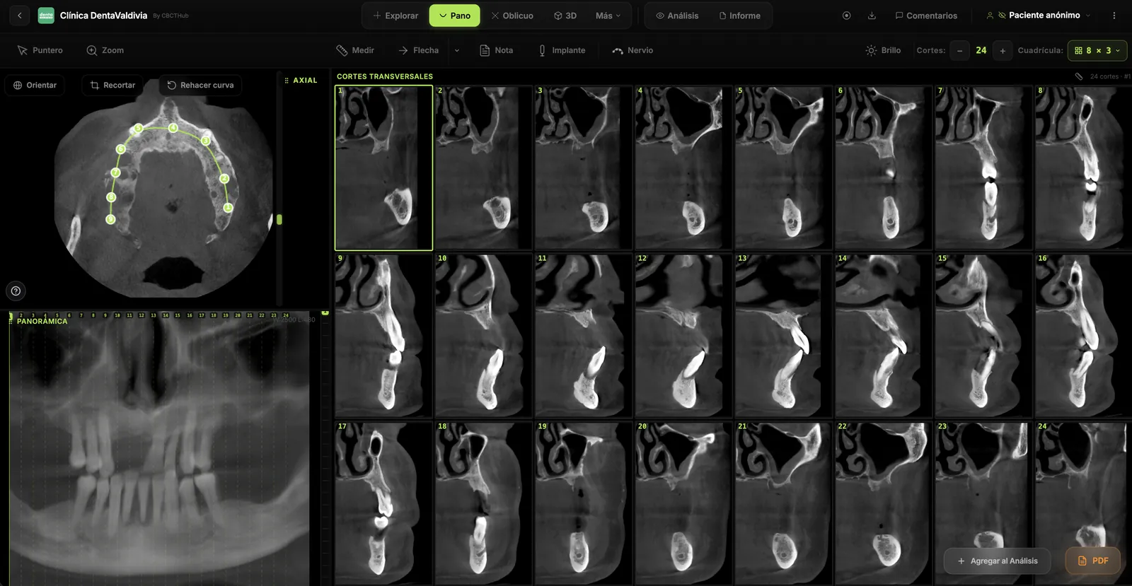
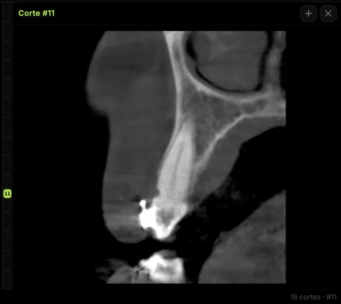
When reviewing a CBCT for endodontic purposes, focus on axial slices at the level of the root apices. Use the scroll function to trace each root from crown to apex. Cross-reference findings in sagittal and coronal views. An online CBCT viewer with synchronized MPR crosshairs makes this workflow fast and intuitive — you can share the scan with the referring dentist so they see exactly what you see.
Try CBCTHub for free
Upload, view, and share DICOM scans in the cloud. Nothing to install.
Create free accountRelated articles
Browser DICOM viewer vs desktop: what works for CBCT in clinic
Open CBCT in the browser or install a desktop viewer? A clinical decision matrix: MPR performance, offline use, Mac/Windows mix, sharing without CDs, and when native software still wins.
How to open a CBCT without installing software: a practical online-viewer guide
You got a CD, ZIP, or link with a CBCT and no manufacturer viewer. How to open the DICOM in the browser, measure in mm, and what to do when it fails.
Mandibular canal and inferior alveolar nerve on CBCT: anatomy, variants, and pre-surgical measurement
Locate the mandibular canal and IAN on CBCT: mental loop, bifid/retromolar canal, measurement checklist, and when to escalate from panoramic (ALADA).