How to Create Professional PDF Reports from CBCT Findings

· Updated

Why CBCT Reports Matter
A CBCT scan without a proper report is only half the picture. Referring dentists rely on structured, clear radiological reports to make treatment decisions. A well-crafted report adds clinical value, demonstrates professionalism, and positions your imaging center as an essential partner in the diagnostic process.
What to Include in a CBCT Report
A comprehensive dental CBCT report should contain:
Header Information
- Patient name, ID, and date of birth
- Date of scan acquisition
- Scanner model and acquisition parameters (FOV, voxel size)
- Referring dentist name and clinic
- Clinical indication for the scan
Findings Section
Organize findings systematically. A common approach:
- Teeth: Individual tooth findings (periapical pathology, root fractures, resorption, anomalies)
- Bone: Alveolar bone levels, bone quality assessment, cortical integrity
- Sinuses: Maxillary sinus mucosa, septa, patency of the ostium
- TMJ: Condylar morphology, joint space, articular eminence (if in the FOV)
- Airway: Pharyngeal airway dimensions and any obstructions (if visible)
- Incidental findings: Any unexpected pathology outside the area of clinical interest
Image Captures
Include representative images that illustrate your key findings:
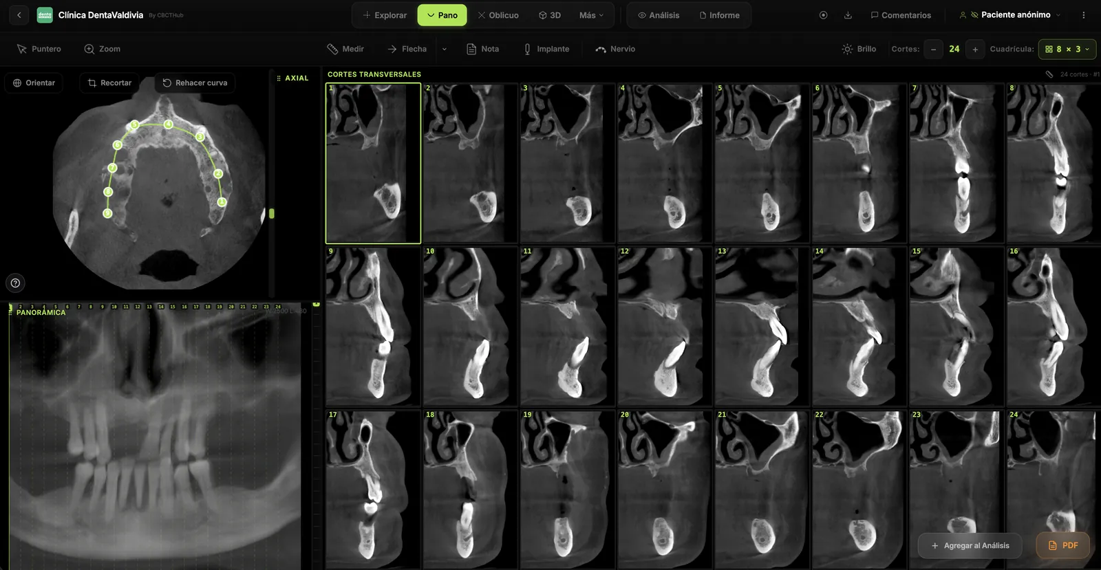
- Annotated axial, sagittal, or coronal slices showing the pathology
- Panoramic reconstruction with arrows or markers
- Cross-sectional views for implant sites with measurements
- Before/after comparisons if follow-up scans are available
Conclusion and Recommendations
Summarize the key findings in plain language. Include clinical recommendations when appropriate — but be careful not to overstep into treatment planning unless that's within your scope.
Formatting Best Practices
- Use your branding: Include your center's logo, colors, and contact information
- Keep it concise: Referring dentists appreciate reports they can scan in under 2 minutes
- Use numbered findings: Makes it easy to reference specific findings in conversation
- Include measurements: Quantitative data (lesion dimensions, bone height, distances to nerve) is more valuable than vague descriptions
- PDF format: Always deliver reports as PDF for universal compatibility and professional appearance
Automating Report Generation
Modern CBCT viewer platforms can streamline reporting. Capture findings directly from the viewer, add annotations, attach clinical notes per finding, and export everything as a branded PDF. This workflow — view, annotate, export — eliminates the need to switch between viewer and word processor, saving significant time per report.
Try CBCTHub for free
Upload, view, and share DICOM scans in the cloud. Nothing to install.
Create free accountRelated articles
Dental CBCT in the cloud: day-to-day storage and sharing in clinic
What “CBCT in the cloud” means in clinic: store the original DICOM, access it from multiple chairs, and share with access control — without mistaking it for a full PACS or a WhatsApp ZIP.
Browser DICOM viewer vs desktop: what works for CBCT in clinic
Open CBCT in the browser or install a desktop viewer? A clinical decision matrix: MPR performance, offline use, Mac/Windows mix, sharing without CDs, and when native software still wins.
Dental DICOM software: a checklist for choosing a CBCT viewer
Before adopting a dental DICOM or CBCT viewer in the clinic: checklist for interoperability, MPR, mm measurements, share without CD, privacy, and TCO.