How to Read a CBCT Scan: A Beginner's Guide for Dentists

· Updated

Getting Started with CBCT Interpretation
Reading a CBCT scan for the first time can feel overwhelming. Unlike traditional 2D radiographs, you're now working with hundreds of slices across three planes. But with a systematic approach, any dentist can learn to interpret CBCT images confidently.
The key is understanding the three standard orientations and developing a consistent review workflow that ensures you never miss critical findings.
The Three Planes of CBCT
Every CBCT volume can be viewed in three orthogonal planes:
- Axial (horizontal): Imagine slicing the head from top to bottom. These views are ideal for evaluating the overall arch shape, cross-sectional bone anatomy, and pathology location relative to buccal and lingual cortices.
- Sagittal (lateral): Slices from left to right, like viewing the patient from the side. Excellent for assessing the relationship between tooth roots, the mandibular canal, and the maxillary sinus floor.
- Coronal (frontal): Slices from front to back. Perfect for evaluating the nasal cavity, sinuses, and bilateral comparison of structures.
Anatomical Landmarks to Identify First
Before focusing on the area of clinical concern, take a moment to identify key landmarks:
- Inferior alveolar nerve canal — appears as a dark tube running through the mandible
- Mental foramen — where the nerve exits the mandible, typically near the premolar region
- Maxillary sinus floor — the boundary between alveolar bone and sinus
- Incisive canal (nasopalatine) — in the anterior maxilla, often mistaken for pathology
- Cortical bone boundaries — buccal and lingual plates that define available bone width
A Systematic Review Workflow
Follow this order every time you review a CBCT scan:
- Step 1: Start with axial slices, scroll through the entire volume top to bottom to check for unexpected findings
- Step 2: Move to the sagittal view and focus on the area of clinical interest
- Step 3: Use the coronal view to confirm findings and assess bilateral symmetry
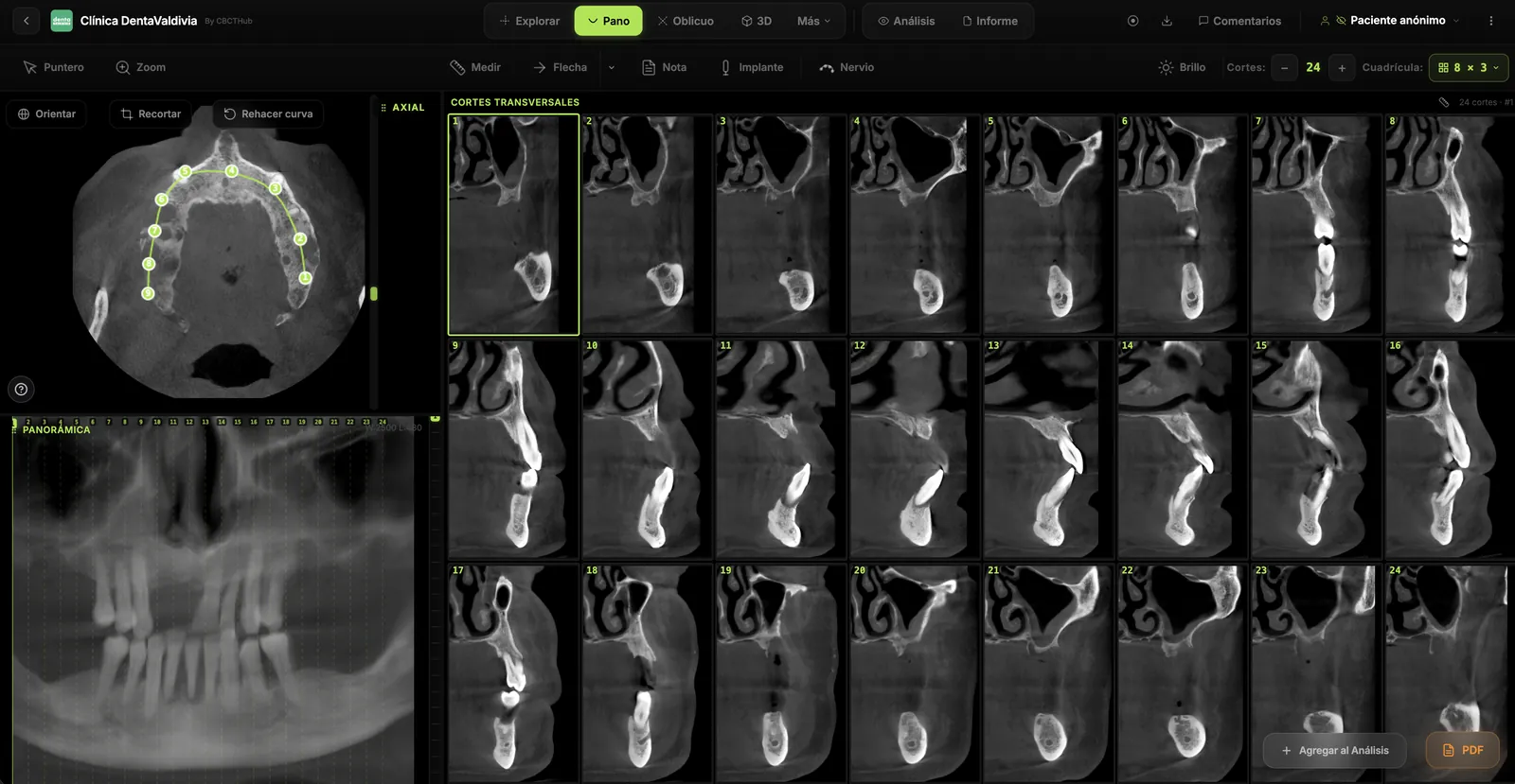
- Step 4: Generate a panoramic reconstruction to see the full arch in context
- Step 5: Use cross-sectional slices for implant planning or detailed bone assessment
Common Mistakes in CBCT Reading
Beginners often fall into these traps:
- Tunnel vision: Only looking at the area of interest and missing incidental findings like sinus pathology or cysts
- Wrong window/level settings: Using bone presets when soft tissue evaluation is needed, or vice versa
- Confusing normal anatomy with pathology: The incisive canal and nutrient canals can mimic periapical lesions in certain views
Practice Makes Perfect
The best way to improve CBCT interpretation is consistent practice. With an online CBCT viewer like CBCTHub, you can access scans from any device, scroll through MPR views, and use measurement tools to build your diagnostic confidence — all without installing desktop software.
Try CBCTHub for free
Upload, view, and share DICOM scans in the cloud. Nothing to install.
Create free accountRelated articles
Endodontic CBCT: AAE/AAOMR 2025 indications
When to justify CBCT in endodontics per the AAE/AAOMR 2025 Position Statement: selective use, limited FOV, ALADAIP, and a practical reading checklist.
Dental CBCT in the cloud: day-to-day storage and sharing in clinic
What “CBCT in the cloud” means in clinic: store the original DICOM, access it from multiple chairs, and share with access control — without mistaking it for a full PACS or a WhatsApp ZIP.
Browser DICOM viewer vs desktop: what works for CBCT in clinic
Open CBCT in the browser or install a desktop viewer? A clinical decision matrix: MPR performance, offline use, Mac/Windows mix, sharing without CDs, and when native software still wins.