CBCT for Supernumerary and Impacted Teeth: 3D Localization Guide

Why Impacted Teeth Need 3D Imaging
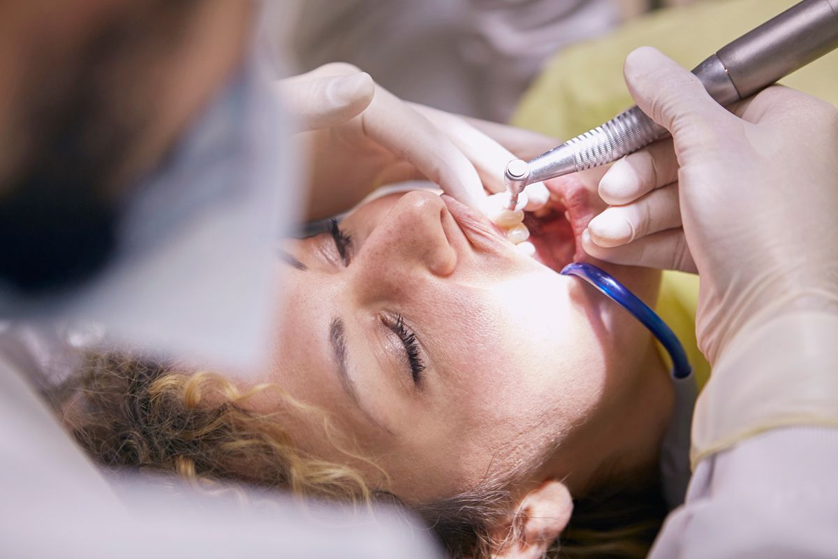
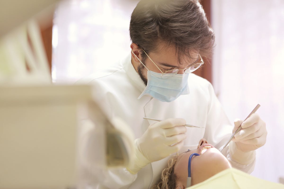
Impacted and supernumerary teeth are among the most common reasons for CBCT referral in dentistry. While panoramic radiographs can detect these teeth, they cannot provide the 3D spatial information needed for safe surgical extraction or orthodontic traction. Questions that 2D imaging cannot reliably answer — Is the impacted canine buccal or palatal? Is the mesiodens inverted? Is there root resorption of adjacent teeth? — are precisely what CBCT excels at answering.
Supernumerary Teeth (Mesiodens and Others)
Supernumerary teeth occur in 1-3% of the population, with the mesiodens (supernumerary tooth in the premaxilla) being the most common. They can cause delayed eruption, displacement, or root resorption of permanent teeth.
CBCT reveals the exact 3D position of the supernumerary tooth: buccal or palatal location, orientation (normal, inverted, or transverse), proximity to and potential resorption of adjacent roots, relationship to the nasal floor and incisive canal, and developmental stage (fully formed vs. developing).
This information directly determines the surgical approach — a palatal approach for palatally positioned teeth vs. a labial approach for buccally positioned ones — reducing surgical time and minimizing risk to adjacent teeth and structures.
Ectopic Maxillary Canines
Ectopic (palatally or buccally displaced) maxillary canines are present in approximately 2% of the population and represent one of the most clinically challenging impactions. The treatment decision — surgical exposure with orthodontic traction vs. extraction — depends on accurate 3D localization.
CBCT assessment of ectopic canines should evaluate the canine's tip position relative to the lateral incisor root (buccal, palatal, or overlapping), angulation of the canine relative to the midline and occlusal plane, distance from the canine tip to the occlusal plane (eruption path length), resorption of lateral incisor roots (present in up to 48% of cases with impacted canines, often invisible on panoramic radiographs), root morphology of the canine itself (dilaceration, incomplete development), and available space in the arch for alignment.
Other Common Impactions
- Impacted premolars: CBCT shows the relationship to deciduous molar roots and the developing premolar position, guiding the decision between extraction and orthodontic management.
- Impacted second molars: CBCT reveals the angulation and proximity to the first molar and IAN canal.
- Odontomas: These benign tumors can block tooth eruption. CBCT localizes them precisely for minimally invasive surgical removal.
CBCT-Guided Surgical Planning
Beyond localization, CBCT enables precise surgical planning: choosing the correct flap design (buccal vs. palatal access), identifying the most direct surgical path with least bone removal, predicting the difficulty of extraction based on root morphology, and planning orthodontic bracket placement and traction direction for canine eruption cases.
Interdisciplinary Communication
Impacted tooth management often involves the orthodontist, oral surgeon, and sometimes the pediatric dentist working as a team. With CBCTHub, the initial CBCT scan can be shared via a link with all team members. The orthodontist plans the space preparation while the surgeon plans the exposure, both reviewing the same 3D images in their browser — ensuring aligned treatment goals before the patient reaches the chair.
Try CBCTHub for free
Upload, view, and share DICOM scans in the cloud. Nothing to install.
Create free accountRelated articles
Mac vs PC for opening CBCT: what to consider before buying
You need to buy a computer for your clinic or imaging center and are torn between Mac and PC. Real criteria for CBCT, no marketing.
iPad for dental radiology: which model is best in 2026
You want to use iPad to review CBCT scans with patient or referrer. Which model fits best? Honest guide based on real clinical use.
How to migrate from CD to cloud without losing old scans
You have years of CBCT scans on CDs and external drives. Here is a step-by-step plan to migrate to cloud without losing a single one.