CBCT for Pre-Prosthetic Assessment and Full-Mouth Rehabilitation

When Prosthetic Dentistry Meets 3D Imaging
Full-mouth rehabilitation is one of the most complex undertakings in dentistry. It requires precise assessment of remaining bone, existing teeth and roots, sinus and nerve anatomy, and the vertical dimension of occlusion. Traditionally planned with panoramic radiographs, periapical series, and mounted casts, modern full-mouth rehabilitation increasingly relies on CBCT imaging for its comprehensive 3D assessment capabilities.
Pre-Prosthetic CBCT Assessment Checklist
When planning complex prosthetic cases, a systematic CBCT evaluation should cover:
- Residual ridge assessment: Height, width, and cross-sectional shape of edentulous ridges. Knife-edge ridges, undercuts, and concavities that affect denture stability or implant placement feasibility.
- Retained roots and pathology: CBCT detects retained root tips, residual cysts, and other pathology that may be invisible on panoramic radiographs — all of which must be addressed before prosthetic treatment.
- Bone quality and quantity: For implant-supported prostheses, CBCT provides detailed bone volume data at every potential implant site, supporting decisions about implant number, size, and position.
- Maxillary sinus: Pneumatization of the maxillary sinus is common in the posterior maxilla after tooth loss. CBCT reveals the available bone height beneath the sinus and determines whether sinus augmentation is needed.
- Mandibular canal: For lower arch rehabilitation, CBCT maps the IAN canal precisely, determining the available bone height for implants in the posterior mandible.
- Mental foramen: Position varies significantly between patients. CBCT localization prevents nerve injury during anterior mandibular implant placement.
- Tori and exostoses: Bony protuberances that may interfere with prosthesis design or require surgical removal.
All-on-4 / All-on-6 Planning with CBCT
The All-on-4 concept — four strategically tilted implants supporting a fixed full-arch prosthesis — requires meticulous CBCT-based planning. Key CBCT assessments include anterior bone volume for the two axial implants, available bone lateral and anterior to the maxillary sinus (upper) or anterior to the mental foramen (lower) for the tilted posterior implants, bone density at each planned site, and angulation calculations for the tilted implants to avoid vital structures while maximizing anterior-posterior spread.
Overdenture Planning
For implant-retained overdentures (typically 2-4 implants), CBCT helps select optimal implant positions that maximize retention while working within available bone. It also reveals interforaminal bone anatomy in the mandible, where the standard two-implant overdenture positions are planned.
Digital Integration
Modern full-mouth rehabilitation workflows integrate CBCT data with digital impressions (intraoral scans) and digital wax-ups to create a fully digital treatment plan. The CBCT provides the bone and anatomy data, the intraoral scan provides the soft tissue and opposing arch data, and the digital wax-up defines the prosthetic outcome — all merged in planning software for implant positioning that is driven by the final prosthetic design.
With CBCTHub, the CBCT component of this workflow becomes instantly shareable. The prosthodontist, surgeon, and dental laboratory can all access the scan via a link, correlating it with the digital wax-up and intraoral scan data to ensure a cohesive treatment plan. This collaborative approach leads to more predictable outcomes and fewer surprises on surgery day.
Try CBCTHub for free
Upload, view, and share DICOM scans in the cloud. Nothing to install.
Create free accountRelated articles
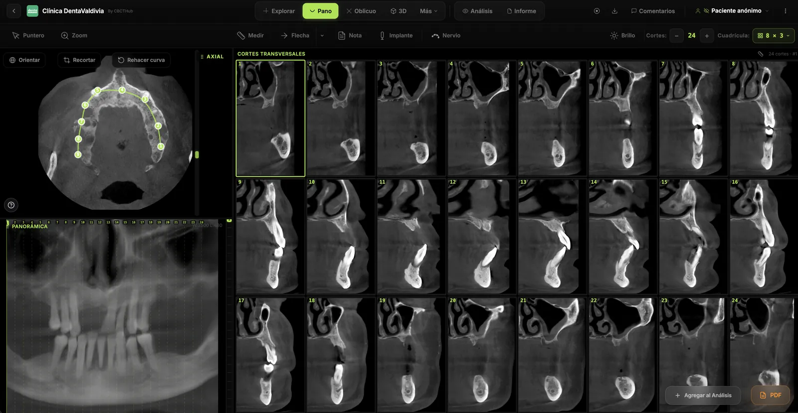
Dental CBCT in the cloud: day-to-day storage and sharing in clinic
What “CBCT in the cloud” means in clinic: store the original DICOM, access it from multiple chairs, and share with access control — without mistaking it for a full PACS or a WhatsApp ZIP.
Browser DICOM viewer vs desktop: what works for CBCT in clinic
Open CBCT in the browser or install a desktop viewer? A clinical decision matrix: MPR performance, offline use, Mac/Windows mix, sharing without CDs, and when native software still wins.
Dental DICOM software: a checklist for choosing a CBCT viewer
Before adopting a dental DICOM or CBCT viewer in the clinic: checklist for interoperability, MPR, mm measurements, share without CD, privacy, and TCO.