The complete CBCT viewer
for diagnosis and treatment planning
Multiplanar reconstruction, panoramic, 3D view, measurements, annotations and signed reports. All in your browser, no install required.
100% in the browser
No downloads, no plugins. Runs in Chrome, Safari, Edge, and Firefox.
Local processing
DICOMs are processed via WebAssembly on your machine. Nothing hits the server.
Any CBCT scanner
Works with Carestream, Planmeca, Vatech, Sirona, Morita, NewTom, and more.
A tool for every job
Everything you need to read a CBCT
Each view designed for a specific clinical case. Switch between them with one click — no annotations or settings lost.
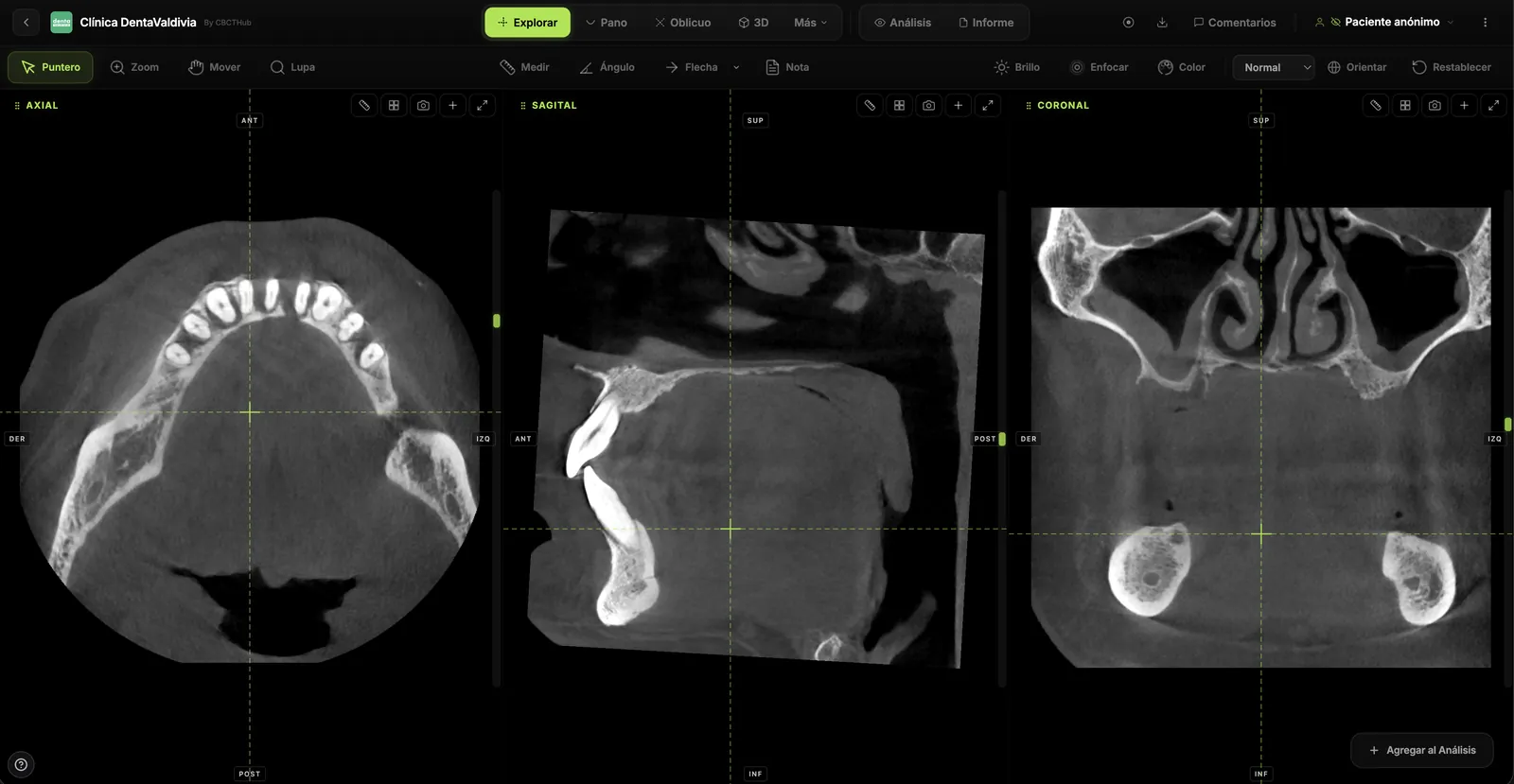
Synced multiplanar reconstruction
The three orthogonal views — axial, sagittal, coronal — synced via crosshair. Move the cursor in one and the others follow the same point. Like any desktop software, but in your browser.
app-explorar.webp
- Dental window/level presets (bone, dental, soft tissue, roots, maxillary sinus, TMJ)
- MIP, MinIP and average modes with live-adjustable slab thickness
- Measurements, angles, arrows and notes that persist across sessions
- Quad and oct view to review multiple slices at once
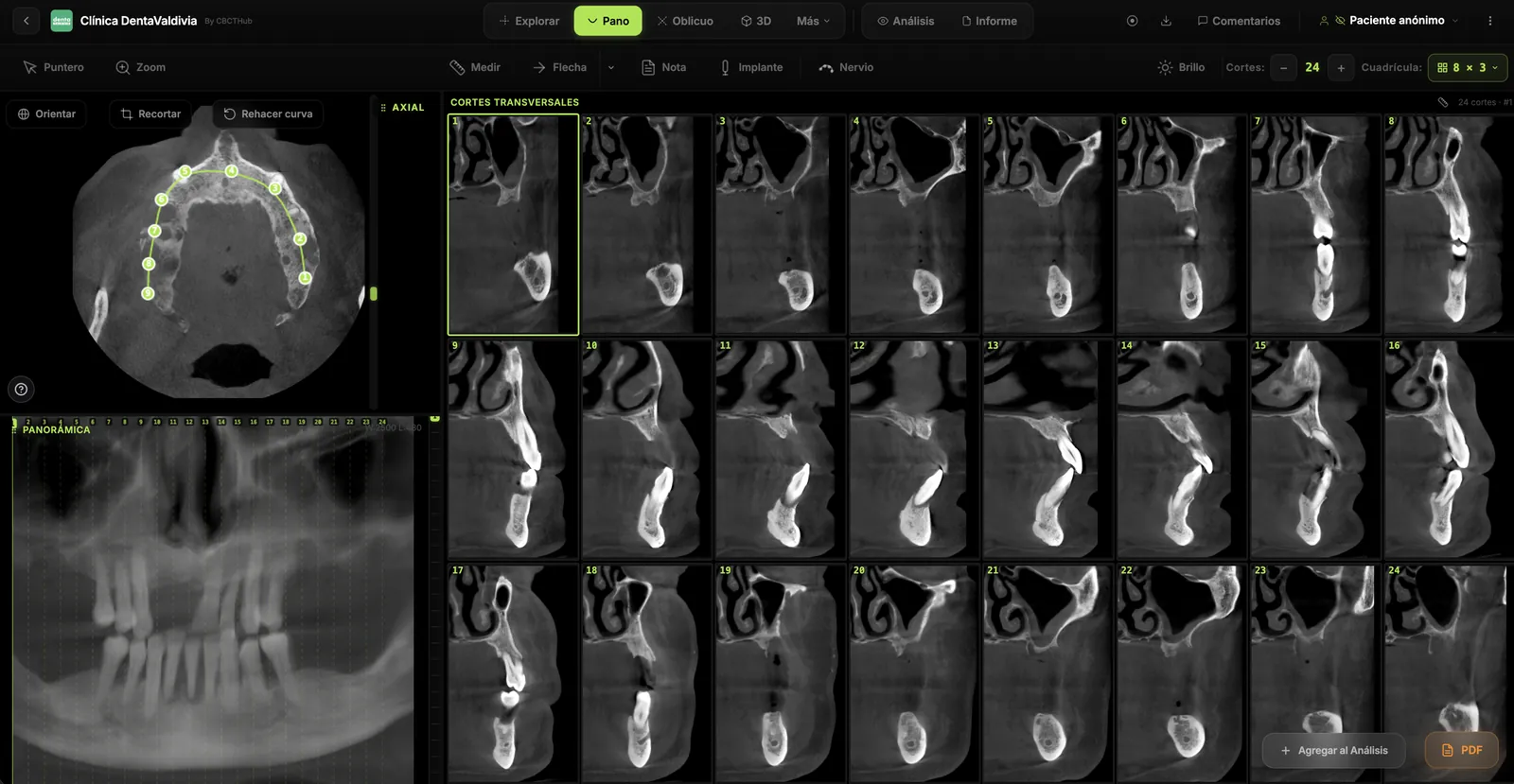
Panoramic reconstructed from the CBCT
Trace the dental arch on the axial slice and the panoramic regenerates instantly, alongside cross-sections with configurable thickness. If the curve isn't right, redraw it in seconds without losing anything.
app-panoramica.webp
- Curve editable point-by-point or redrawn from scratch anytime
- Panoramic and cross-section thickness adjustable in millimeters
- Virtual implants with configurable diameter, length and rotation
- Inferior alveolar nerve tracing, left and right
app-vista3d-full.webp
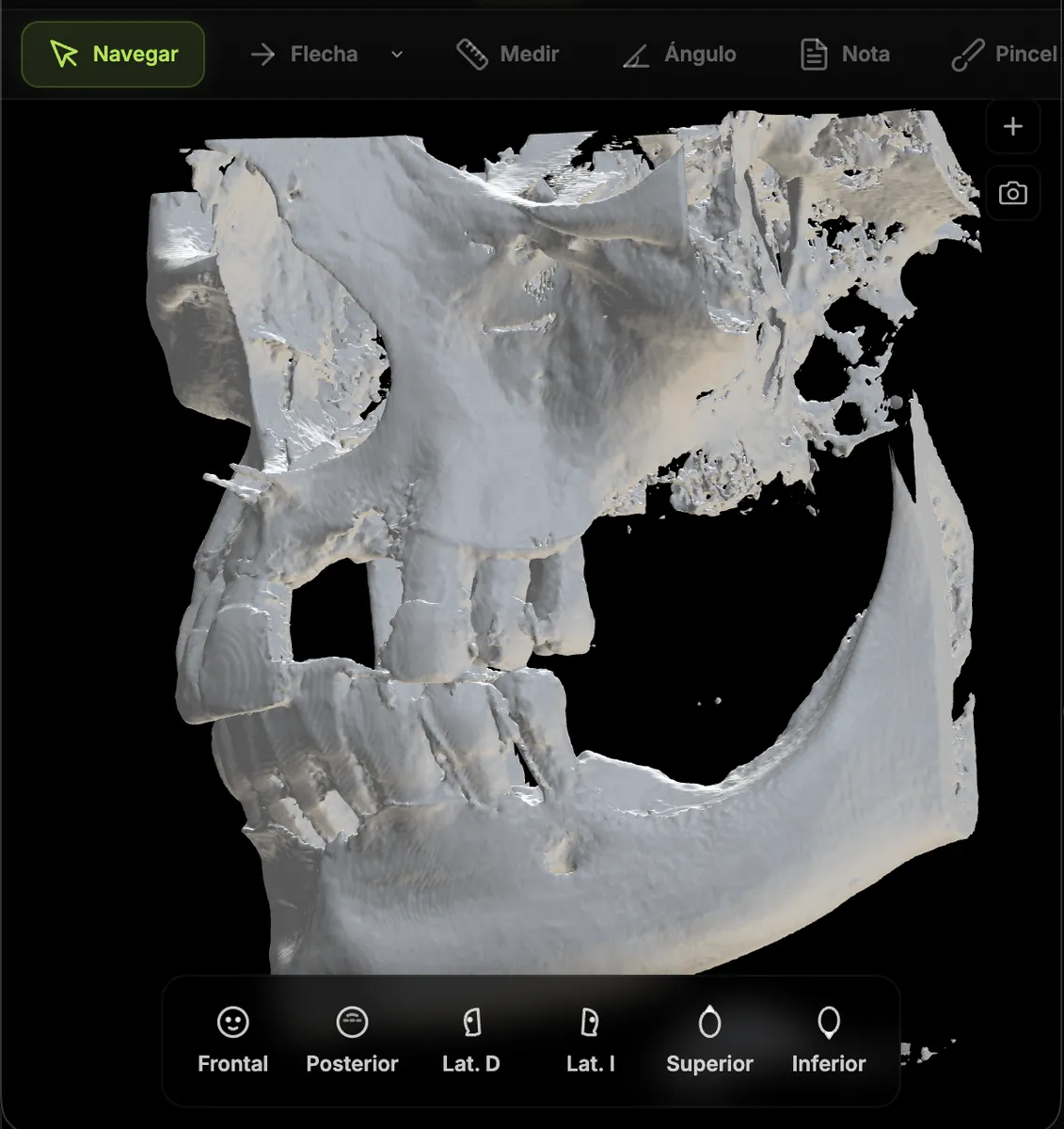
Volume and surface in real time
Classic volume rendering for an overview, plus iso-surface reconstruction with Marching Cubes for a clean look like surgical software. Rotate, crop, export — all in the browser.
- WebGL render with trackball rotation, scroll zoom and smooth pan
- X / Y / Z clipping planes to slice the model and look inside
- STL export ready for 3D printing or CAD software
- High-res capture, 3D-anchored annotations and presentation mode
app-oblicuo-full.webp
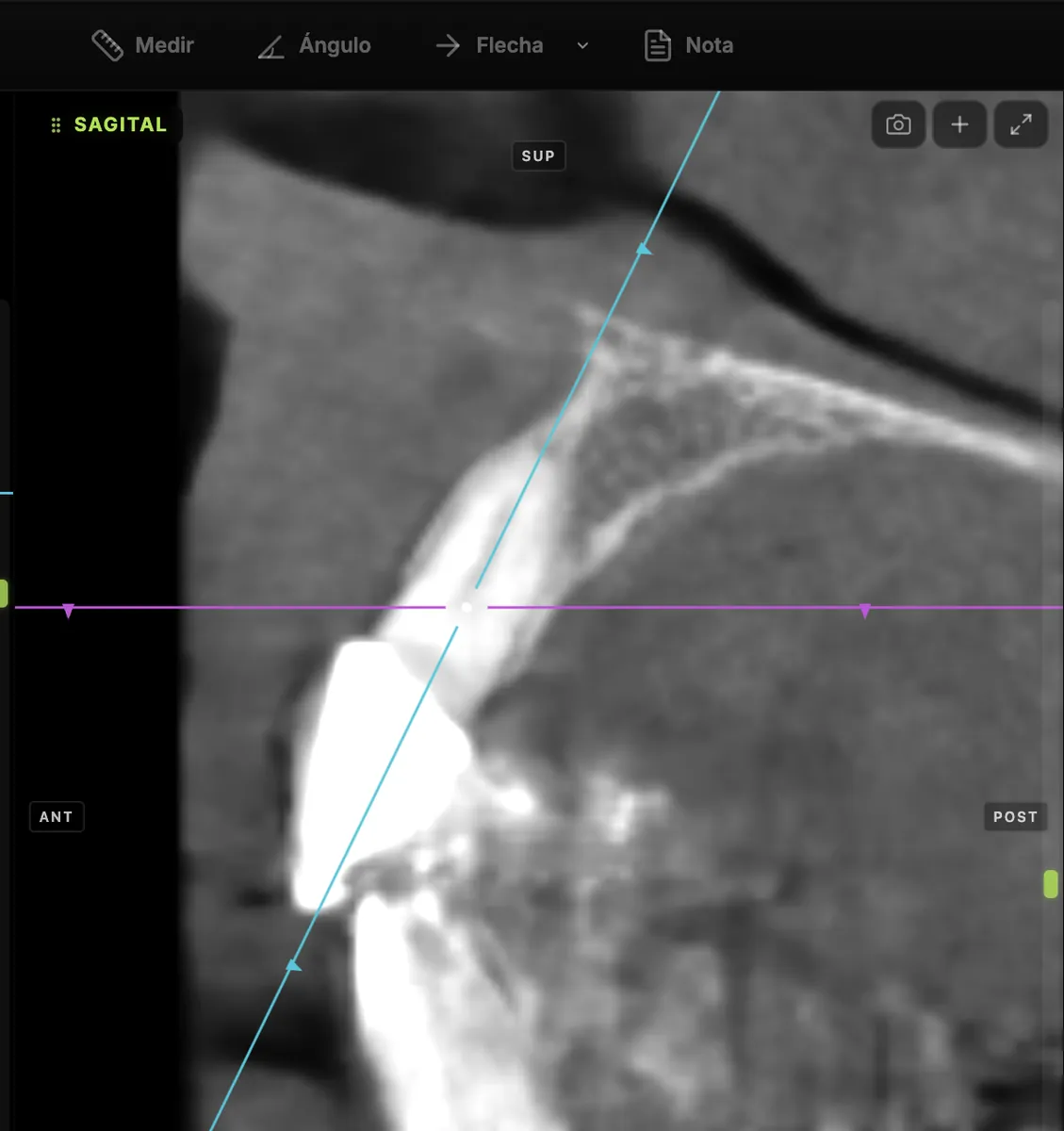
Slices in any orientation
When orthogonal planes aren't enough, the oblique view lets you see the volume from the exact angle you need — an implant axis, an angled root, a facial asymmetry.
- Fully rotatable crosshair across all three axes
- Independent slab thickness for smoothing or detail
- Persistent configuration across sessions for the same exam
Temporomandibular joint
Split-screen with right and left TMJ side by side. Trace the condylar axis and get automatic perpendicular slices to evaluate the joint with the precision the case requires.
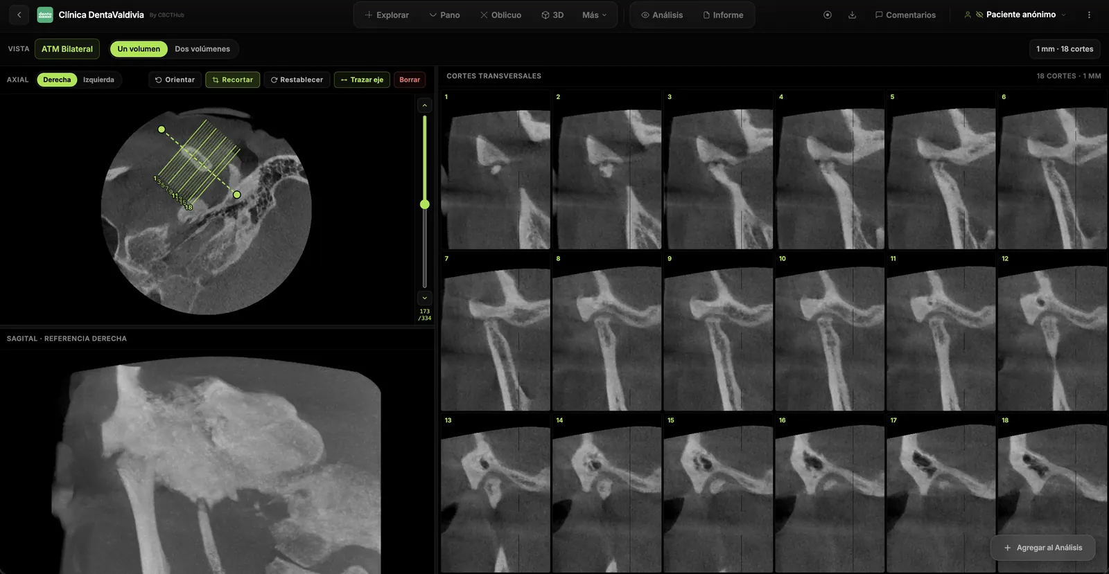
app-atm.webp
- Bilateral comparison right vs. left on screen
- Condylar axis tracing with automatic perpendicular slices
- Support for cases with two volumes (closed and open) side by side
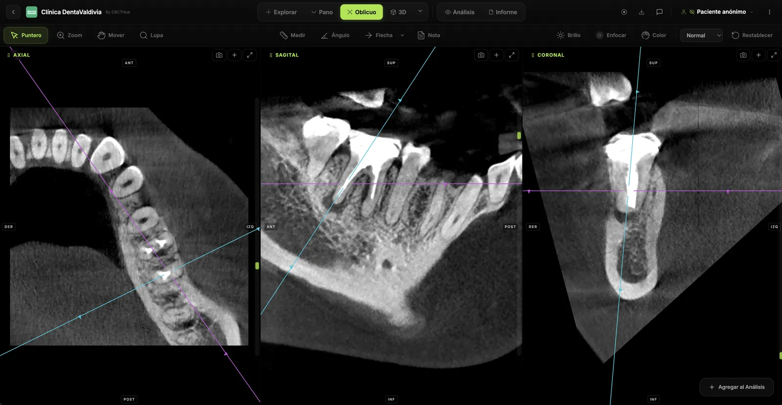
esp-endodoncia.webp
From finding to signed report
Measurements in millimeters, arrows, circles, notes, angles. All saved automatically. When you're done, generate a radiology PDF report with your logo, digital signature and a verification QR code.
- Real 3D measurements in mm on any slice or view
- Anchored annotations that follow the model when you rotate the 3D view
- Radiology PDF report with template, logo and professional signature
- Anonymous public link to share with the referring dentist
Use cases
Built for every specialty
The right tools configured for your discipline, saving you the learning curve of generic software.
app-panoramica.webp
Implantology
Measure available bone height and width, evaluate distance to the inferior alveolar nerve and maxillary sinus, plan with virtual implants on the panoramic and cross-sections.
esp-endodoncia.webp
Endodontics
Identify accessory canals, periapical lesions and root curvatures with oblique slices at the exact angle of each root. Precise measurements for working length.
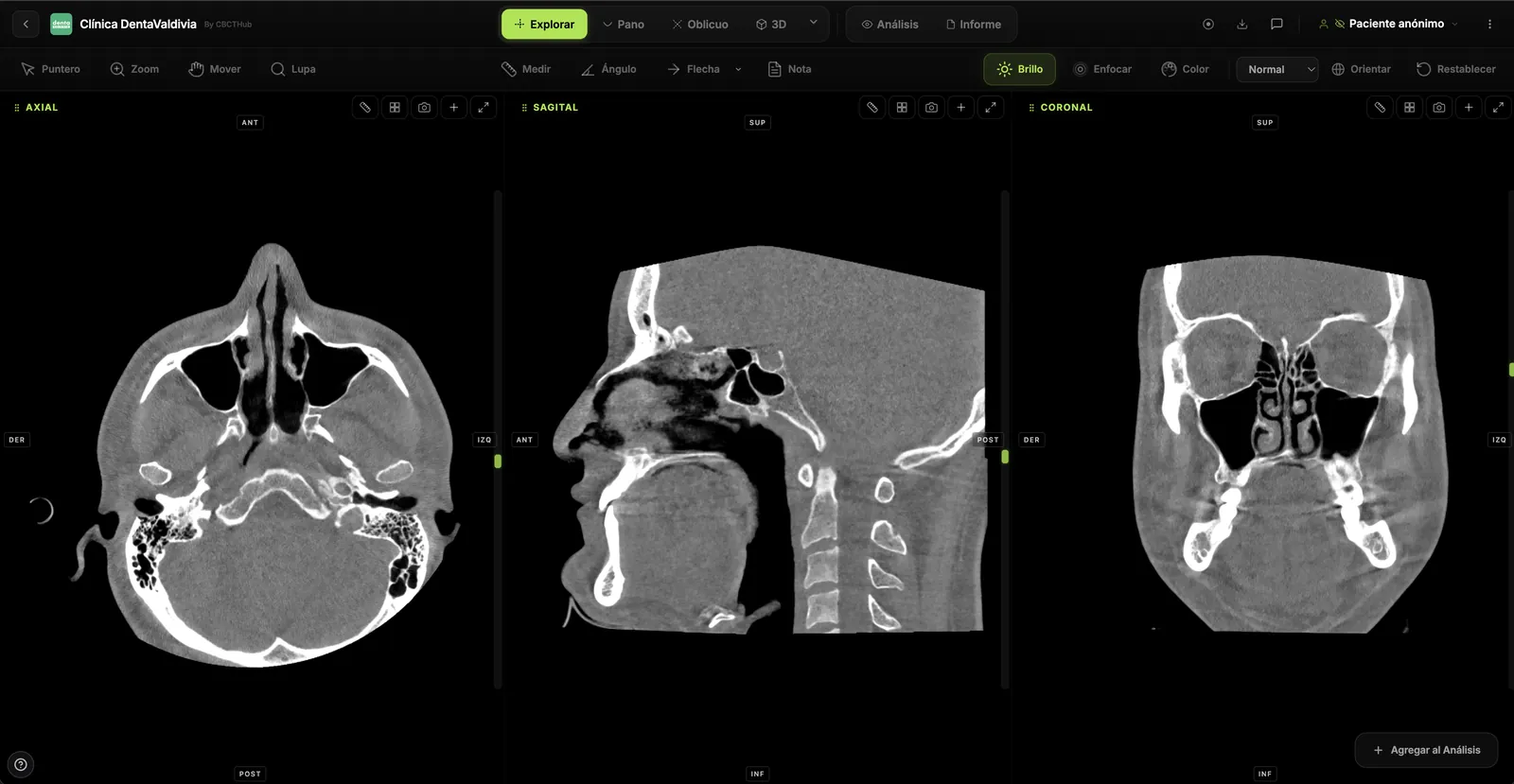
esp-ortodoncia.webp
Orthodontics & orthognathics
Cephalometric analysis, third-molar evaluation, facial asymmetries, condylar position. 3D view and STL export for virtual articulators or printed models.
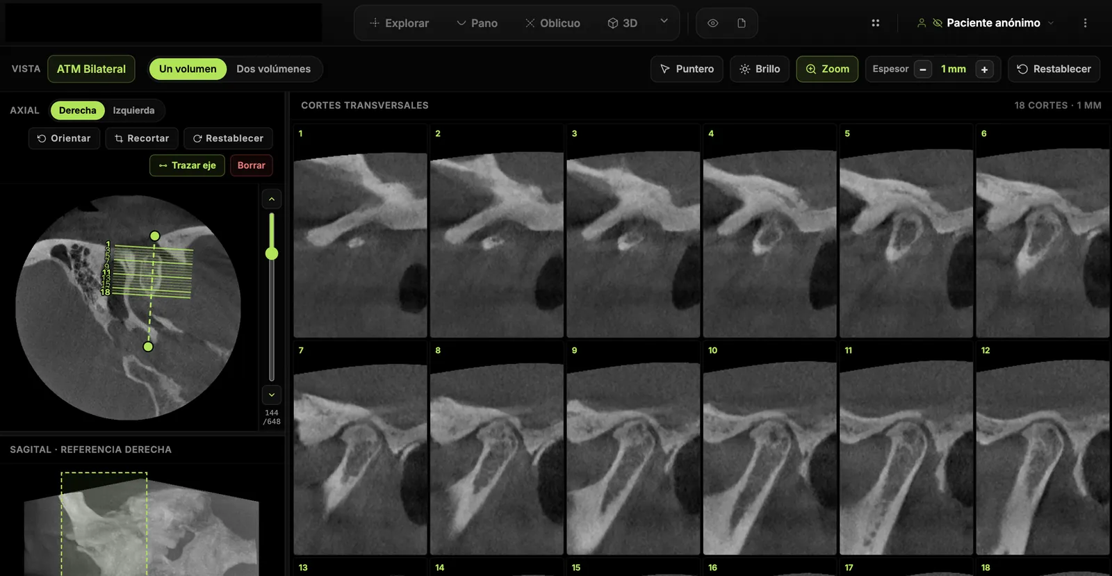
esp-atm.webp
TMJ dysfunction
Right vs. left comparison, joint space evaluation, condylar position and head morphology. Support for closed- and open-mouth cases.
Built for your workflow
Made for how you actually work
The technical decisions that matter, made for you.
No install required
Runs in Chrome, Safari, Edge and Firefox. No plugins, no updates, no IT permissions. Open your browser and you're in.
Local processing
The CBCT is reconstructed via WebAssembly on your computer. It doesn't go to the server to be processed — patient privacy stays on your machine.
Share with a link
Send a link to the referring dentist. They open it in their browser and see the full viewer, no download required. No CDs, no USBs, no asking them to install software.
Three languages
Full interface in Spanish, English and Portuguese. Switch with one click — reports and emails are also generated in the recipient's language.
Any brand
Carestream, Planmeca, Vatech, Sirona, Morita, NewTom, Acteon, KaVo and more. If your scanner exports standard DICOM, CBCTHub reads it.
Your brand, your report
Your clinic logo, header and digital signature. The patient receives a PDF with your identity, not ours.
Compatibility
Works with every CBCT brand
If your scanner exports standard DICOM, it works with CBCTHub.
Don't see your brand? View full list →
Frequently asked
Common questions
Start using CBCTHub today
Create your free account and open your first CBCT in under a minute. No credit card, no commitment.
Free plan included forever. No credit card required.