CBCT Radiation Dose in Dentistry: What Patients and Clinicians Should Know

Understanding Radiation Dose in Dental CBCT
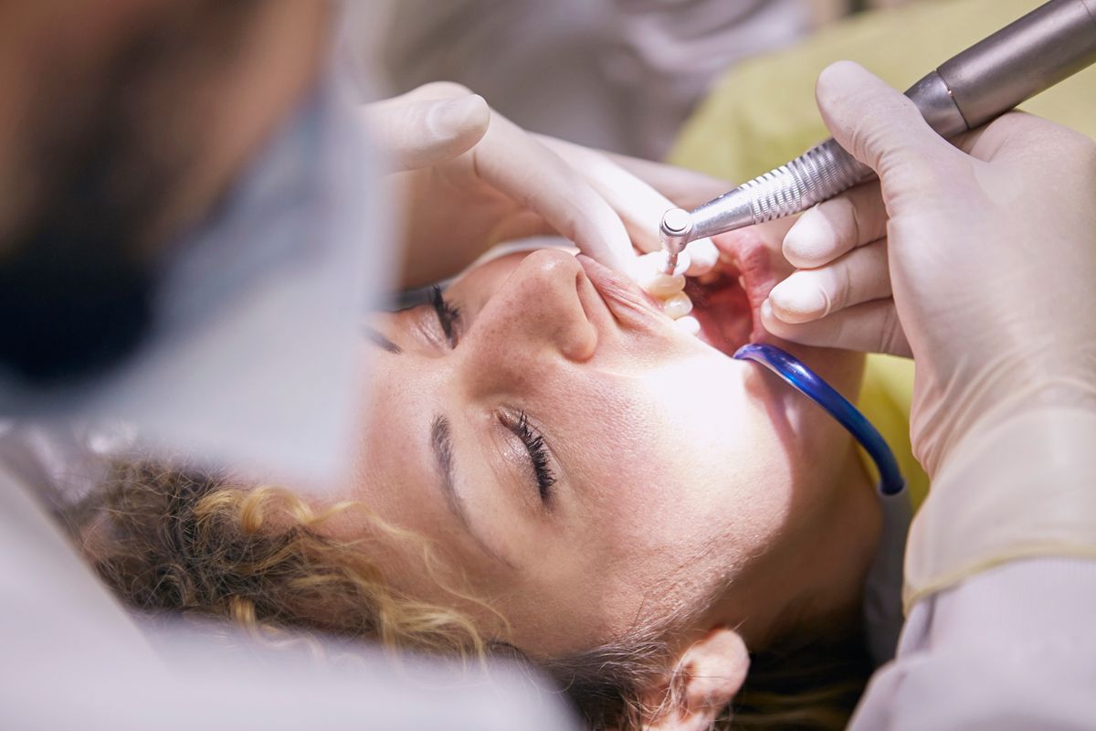
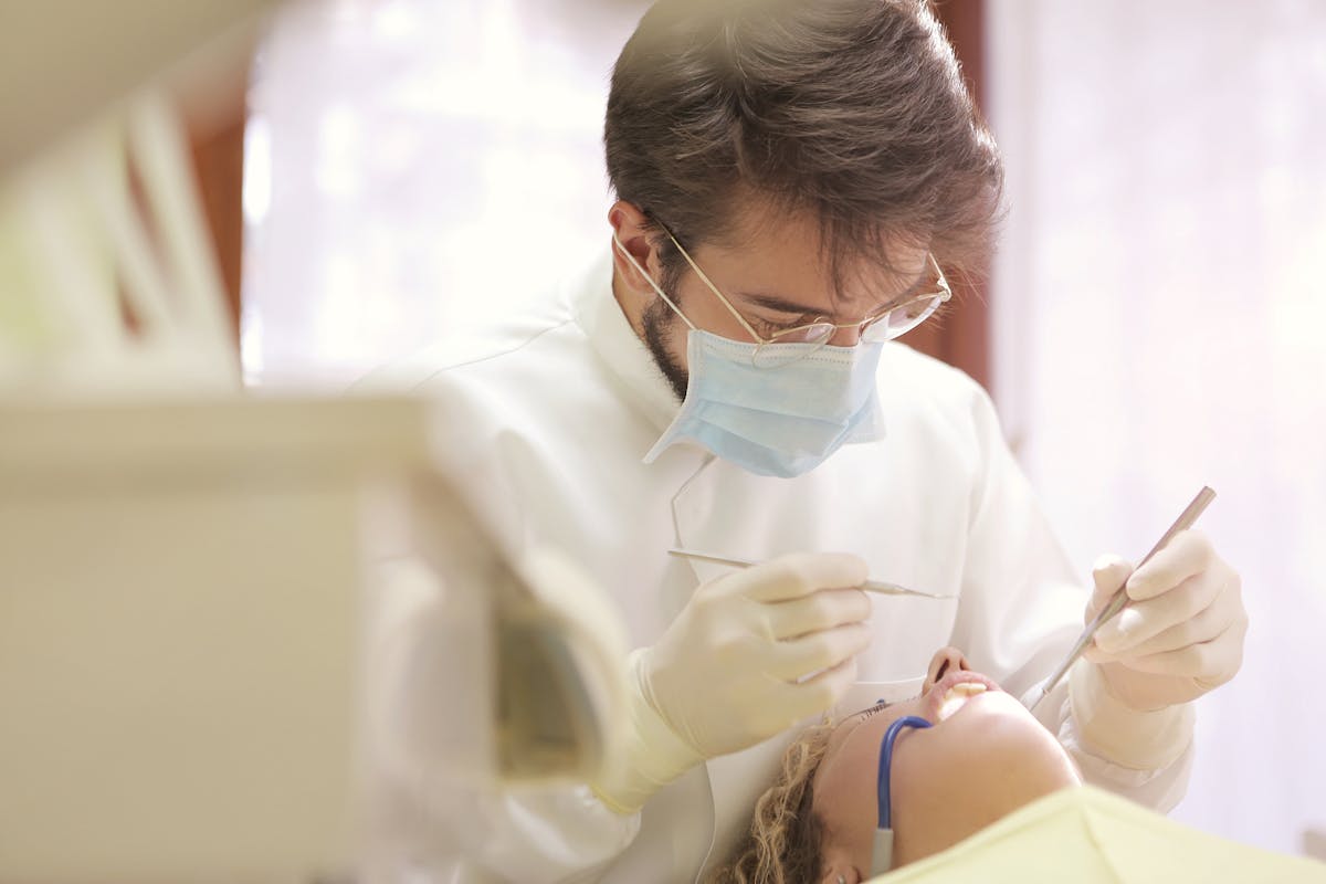
One of the most common concerns patients have about CBCT imaging is radiation exposure. As clinicians, it's our responsibility to understand the actual doses involved, how they compare to other imaging modalities, and how to apply the ALARA principle (As Low As Reasonably Achievable) in practice.
How CBCT Dose Compares
Radiation dose is measured in microsieverts (μSv). Here's how dental CBCT compares to other common exposures:
- Single periapical radiograph: 5-10 μSv
- Panoramic radiograph: 10-25 μSv
- Full-mouth series (18 films): 90-180 μSv
- Dental CBCT (small field): 20-100 μSv
- Dental CBCT (large field): 50-200 μSv
- Medical CT of the head: 1,500-2,000 μSv
- Annual background radiation: ~3,000 μSv
A small-field dental CBCT scan delivers roughly the same dose as a full-mouth series of periapical radiographs, and 10-100 times less than a medical CT scan.
Factors That Affect CBCT Dose
Several factors influence the radiation dose from a CBCT scan:
- Field of view (FOV): Smaller FOV = lower dose. Use the smallest FOV that covers the area of clinical interest.
- Resolution settings: Higher resolution means more radiation. Standard resolution is adequate for most clinical needs.
- Exposure parameters: kVp and mA settings directly affect dose. Use manufacturer-recommended protocols.
- Patient size: Pediatric patients should use reduced exposure settings.
ALARA in Practice
Following ALARA principles for dental CBCT means:
- Justification: Only order a CBCT when the diagnostic question cannot be answered by lower-dose imaging (periapical, panoramic)
- Optimization: Use the smallest FOV, lowest acceptable resolution, and appropriate exposure settings for each case
- Documentation: Record the clinical indication for every CBCT scan ordered
Communicating with Patients
When patients ask about radiation, provide context. A dental CBCT scan typically delivers less radiation than a day of natural background exposure. The diagnostic information gained — precise nerve location, hidden pathology, accurate measurements for implant planning — directly improves treatment outcomes and safety. The risk of missing a diagnosis without CBCT often outweighs the minimal radiation risk.
Try CBCTHub for free
Upload, view, and share DICOM scans in the cloud. Nothing to install.
Create free accountRelated articles
Mac vs PC for opening CBCT: what to consider before buying
You need to buy a computer for your clinic or imaging center and are torn between Mac and PC. Real criteria for CBCT, no marketing.
How to choose a dental PACS for your clinic or imaging center
A practical checklist of the 10 criteria that matter when choosing a dental PACS: price, storage, viewer, integrations, support, compliance and more.
iPad for dental radiology: which model is best in 2026
You want to use iPad to review CBCT scans with patient or referrer. Which model fits best? Honest guide based on real clinical use.