CBCT Artifacts Explained: Types, Causes, and How to Fix Them
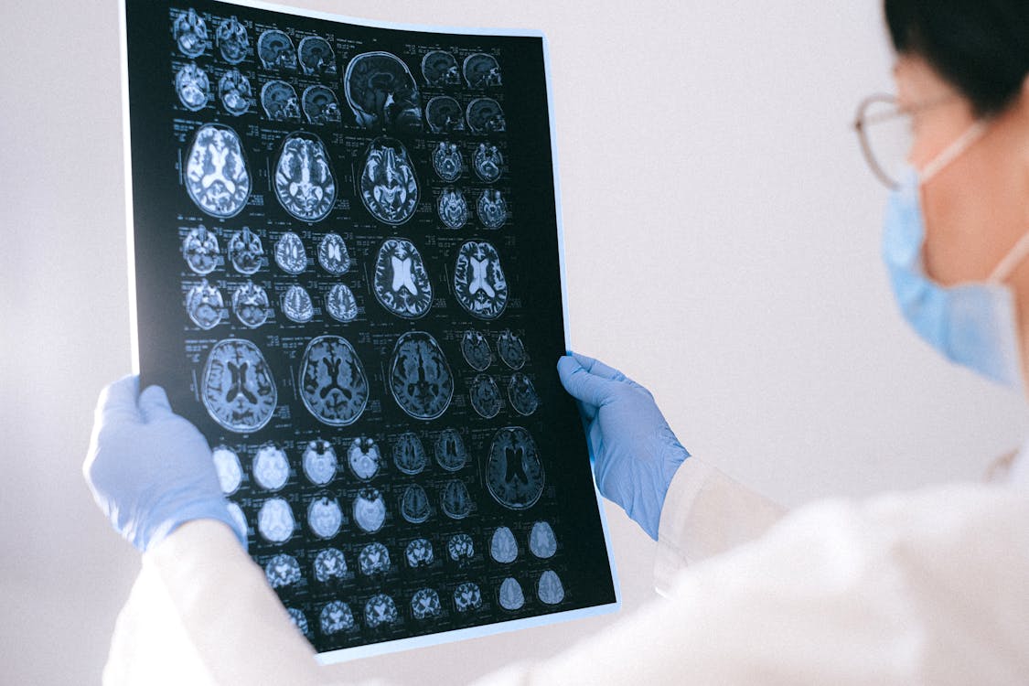
What Are CBCT Artifacts?
Artifacts are image distortions or errors that don't represent actual anatomical structures. In CBCT imaging, artifacts can obscure diagnostic information, mimic pathology, or reduce image quality to the point where the scan becomes non-diagnostic. Understanding what causes artifacts — and how to prevent or minimize them — is essential for every clinician who works with CBCT.
Beam Hardening Artifacts
What it looks like: Dark bands or streaks between dense structures (like between implants or between enamel surfaces). Also appears as a cupping effect where the center of a dense object appears darker than its edges.
What causes it: CBCT uses a polychromatic X-ray beam. As the beam passes through dense material, lower-energy photons are absorbed preferentially, leaving higher-energy photons — this "hardens" the beam. The reconstruction algorithm doesn't account for this spectral shift, creating false attenuation values.
How to minimize it: Beam hardening correction algorithms in modern CBCT software can reduce this artifact. Patient positioning to avoid scanning through the densest path (e.g., tilting the head slightly) may help. When planning implant sites near existing implants, be aware that the area between implants will have degraded image quality.
Metal (Scatter) Artifacts
What it looks like: Bright streaks, starbursts, or white bands radiating from metal objects — crowns, implants, amalgam restorations, orthodontic brackets, and endodontic posts.
What causes it: High-density metals cause severe photon absorption and scatter. The reconstruction algorithm cannot accurately compute attenuation values in the path of these scattered photons, creating streaks in the image.
How to minimize it: Remove all removable metal objects before scanning (earrings, glasses, removable prostheses). Use metal artifact reduction (MAR) algorithms available in many CBCT units. For orthodontic patients, consider scanning before bracket placement when possible. Some software solutions use iterative reconstruction to reduce metal artifacts post-acquisition.
Motion Artifacts
What it looks like: Blurred or doubled contours, especially at sharp edges like cortical bone margins. The entire image may appear "smeared" or lack sharpness.
What causes it: Patient movement during the scan. Even slight movement (1-2 mm) during the 10-40 second acquisition can significantly degrade image quality. Common culprits: swallowing, breathing, involuntary head movement, or patient anxiety.
How to minimize it: Use proper head stabilization with chin rests, head straps, and bite blocks. Instruct the patient clearly before scanning — emphasize staying still and breathing through the nose. Use the shortest scan time that provides adequate image quality. For anxious or pediatric patients, consider a practice run without radiation.
Truncation (Cone-Cut) Artifacts
What it looks like: The structure of interest is partially cut off at the edge of the image, with bright or dark lines at the truncation boundary.
What causes it: The object being scanned extends beyond the selected field of view (FOV). The reconstruction algorithm lacks complete data for voxels at the edge, creating artifacts.
How to minimize it: Select an appropriate FOV that fully encompasses the area of interest with a margin. If the mandible's inferior border is cut off, the scan should be repeated with proper positioning or a larger FOV.
Ring Artifacts
What it looks like: Circular rings or arcs centered on the rotation axis, visible in axial slices.
What causes it: Defective or poorly calibrated detector elements that produce consistently incorrect readings at specific positions.
How to minimize it: Regular flat-field calibration of the CBCT unit per manufacturer's recommendations. If ring artifacts appear suddenly, contact the manufacturer's service team for detector recalibration.
Best Practices for Artifact-Free Scans
Consistent patient preparation and proper technique prevent most artifacts. Remove all metallic objects from the scan area, position the patient carefully using laser guides, select the smallest appropriate FOV and the correct exposure protocol, ensure the patient is stable and comfortable before starting, and keep the CBCT unit calibrated and maintained. When reviewing scans in CBCTHub's online viewer, adjusting window/level settings can sometimes help visualize anatomy that is partially obscured by artifacts.
Try CBCTHub for free
Upload, view, and share DICOM scans in the cloud. Nothing to install.
Create free accountRelated articles

What Is CBCT and Why Every Dental Practice Needs It
Learn how cone beam computed tomography revolutionizes dental imaging and diagnosis. Discover why CBCT is essential for modern dental practices.
How to Read a CBCT Scan: A Beginner's Guide for Dentists
New to CBCT interpretation? Learn how to navigate axial, sagittal, and coronal views, identify key anatomical landmarks, and avoid common reading mistakes.
CBCT for TMJ Disorders: Diagnosis and Treatment Planning
Learn how cone beam CT helps diagnose temporomandibular joint disorders, evaluate condylar morphology, and plan treatment for TMD patients.