Complete Guide to the DICOM Format for Dental Professionals

What Is DICOM?
DICOM (Digital Imaging and Communications in Medicine) is the international standard for storing, transmitting, and displaying medical images. For dental professionals working with digital imaging, understanding the DICOM format is essential for effective data management, sharing, and compliance.
Developed by the American College of Radiology and the National Electrical Manufacturers Association, DICOM ensures that medical images from different manufacturers and systems can be viewed, processed, and shared seamlessly.
DICOM File Structure and Components
A DICOM file consists of:
- File Preamble: A 128-byte header and "DICM" magic word that identifies it as a DICOM file.
- File Meta Information: Contains metadata about the image, including transfer syntax and media storage.
- Dataset: The actual image data and associated attributes organized in a hierarchical structure of tags.
Key DICOM Attributes and Tags
DICOM uses a tag system to organize information. Common tags include:
- Patient Information (0x0010): Patient ID, name, date of birth, sex
- Study Information (0x0020): Study date, study time, study description
- Series Information: Series number, modality (CT, MR, etc.)
- Image Information: Pixel data, image dimensions, image position
- Equipment Information: Manufacturer, model, software version
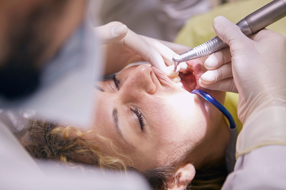
DICOM Modalities in Dental Imaging
Different imaging modalities produce DICOM files with specific characteristics:
CT (Computed Tomography): Volumetric 3D imaging, typical of CBCT systems. CT files contain multiple slices representing different positions through the scanned volume.
CR (Computed Radiography): Digital radiographs from phosphor plate systems.
DX (Intraoral Radiography): Digital intraoral X-ray images.
DICOM Transfer Syntax
Transfer syntax defines how pixel data is encoded within the DICOM file. Common dental imaging transfer syntaxes include:
- Explicit VR Little Endian: Explicit Value Representation with little-endian byte ordering (most common)
- JPEG Lossless: Compressed without loss of diagnostic information
- JPEG 2000 Lossless: Advanced compression maintaining full diagnostic quality
Window/Level (Grayscale Rendering)
CBCT images are acquired as 12-bit or 14-bit grayscale data. Window (width) and level (center) settings control how this data is displayed, allowing clinicians to optimize visualization of different tissues—bone, soft tissue, or air.
DICOM Networking and Protocols
DICOM files are designed for network transmission using the DICOM protocol. This enables:
- DICOM PACS (Picture Archiving and Communication Systems) integration
- Query and retrieve operations across networked systems
- Automated worklist management
- Real-time image streaming
Privacy and De-identification
DICOM includes mechanisms for de-identifying patient data by removing or replacing protected health information. When sharing images for educational or research purposes, proper de-identification is essential for HIPAA compliance.
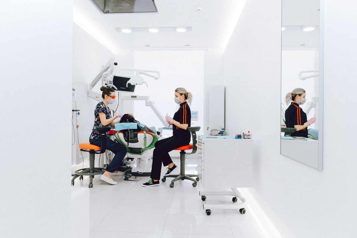
Working with DICOM in Modern Workflows
Contemporary dental practices typically interact with DICOM through specialized viewer software. Understanding basic DICOM concepts helps when troubleshooting compatibility issues, optimizing image quality, or implementing new imaging equipment and software systems.
Conclusion
The DICOM standard is the backbone of digital medical imaging. While the technical details are managed by software, understanding key DICOM concepts—file structure, metadata, transfer syntax, and networking—ensures you can effectively manage dental imaging data and integrate systems across your practice or radiology center.
Try CBCTHub for free
Upload, view, and share DICOM scans in the cloud. Nothing to install.
Create free accountRelated articles
Mac vs PC for opening CBCT: what to consider before buying
You need to buy a computer for your clinic or imaging center and are torn between Mac and PC. Real criteria for CBCT, no marketing.
How to choose a dental PACS for your clinic or imaging center
A practical checklist of the 10 criteria that matter when choosing a dental PACS: price, storage, viewer, integrations, support, compliance and more.

iPad for dental radiology: which model is best in 2026
You want to use iPad to review CBCT scans with patient or referrer. Which model fits best? Honest guide based on real clinical use.