CBCT in Oral Pathology: Detecting Cysts, Tumors, and Bone Lesions
The Role of CBCT in Oral Pathology
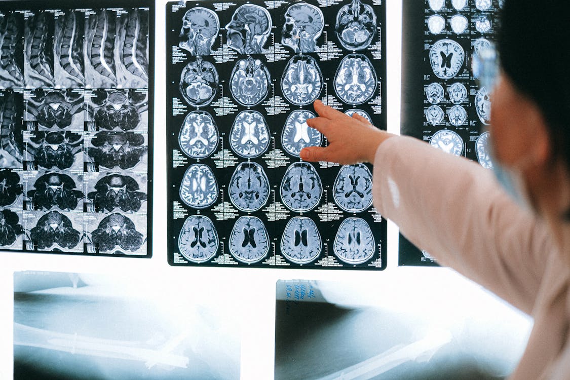
Pathological lesions of the jaws — from common cysts to rare tumors — require accurate assessment of size, location, relationship to vital structures, and effect on adjacent teeth. While panoramic radiographs can detect many lesions, they cannot reveal the 3D extent that determines surgical approach and prognosis.
CBCT provides critical information that 2D imaging cannot: true lesion volume, buccal/lingual cortical involvement, proximity to the inferior alveolar nerve in all planes, tooth root resorption patterns, and internal structure (unilocular vs. multilocular).
Common Odontogenic Cysts on CBCT
Dentigerous (Follicular) Cyst
The second most common odontogenic cyst, always associated with an unerupted tooth crown. On CBCT, it appears as a well-defined, unilocular radiolucency attached at the cementoenamel junction of the unerupted tooth. CBCT reveals the precise relationship to the mandibular canal, sinus floor, and adjacent roots — determining whether marsupialization or enucleation is appropriate.
Radicular (Periapical) Cyst
Associated with a nonvital tooth, appearing as a round radiolucency at the apex. CBCT helps differentiate between a periapical cyst and a granuloma (though histological confirmation is definitive) and reveals whether the lesion has expanded into the sinus or eroded cortical bone.
Odontogenic Keratocyst (OKC)
A clinically significant lesion with a high recurrence rate (25-60%). On CBCT, OKCs appear as well-defined, sometimes scalloped radiolucencies that tend to grow along the medullary space without causing significant expansion. They may be unilocular or multilocular. CBCT is crucial for surgical planning because OKCs can be extensive, extending along the body or ramus of the mandible while maintaining thin cortical walls.
Benign Odontogenic Tumors
Ameloblastoma
The most clinically significant benign odontogenic tumor due to its locally aggressive behavior. On CBCT, ameloblastomas typically appear as multilocular radiolucencies with a "soap bubble" or "honeycomb" pattern. CBCT reveals the true extent of the lesion, cortical expansion or perforation, tooth root resorption, and relationship to the inferior alveolar canal — all critical for planning the extent of surgical resection.
Odontoma
The most common odontogenic tumor, often discovered as an incidental finding or when investigating a delayed tooth eruption. Compound odontomas contain multiple small tooth-like structures (denticles), while complex odontomas appear as an amorphous radiopaque mass. CBCT helps localize the lesion precisely and plan the least invasive surgical approach.
Non-Odontogenic Lesions
CBCT can also characterize non-odontogenic bone lesions such as central giant cell granulomas (multilocular, often in the anterior mandible), aneurysmal bone cysts (expansile, sometimes with internal septations), fibrous dysplasia (ground-glass opacity with altered bone architecture), and Stafne bone defect (lingual mandibular depression — CBCT confirms it's a cortical depression, not a true cyst, avoiding unnecessary surgery).
CBCT's Impact on Treatment Planning
For pathological lesions, CBCT changes the treatment approach in several ways: it determines whether a conservative (enucleation/marsupialization) or aggressive (resection) approach is needed, identifies safe surgical margins by showing the lesion's 3D boundaries, reveals whether reconstruction (bone graft, plate) will be needed, and helps predict the risk of nerve injury during surgery.
When a complex pathology case requires interdisciplinary consultation — between the general dentist, oral surgeon, and pathologist — CBCTHub enables instant scan sharing so every team member can evaluate the 3D anatomy independently and contribute to the treatment plan.
Try CBCTHub for free
Upload, view, and share DICOM scans in the cloud. Nothing to install.
Create free accountRelated articles
Mac vs PC for opening CBCT: what to consider before buying
You need to buy a computer for your clinic or imaging center and are torn between Mac and PC. Real criteria for CBCT, no marketing.
iPad for dental radiology: which model is best in 2026
You want to use iPad to review CBCT scans with patient or referrer. Which model fits best? Honest guide based on real clinical use.
How to migrate from CD to cloud without losing old scans
You have years of CBCT scans on CDs and external drives. Here is a step-by-step plan to migrate to cloud without losing a single one.