CBCT-Guided Implant Surgery: From Digital Planning to Surgical Guides

The Digital Implant Planning Revolution
Dental implant placement has evolved from a purely surgical skill into a digitally planned, prosthetically driven procedure. At the center of this revolution is CBCT imaging, which provides the 3D bone data essential for virtual implant positioning, surgical guide fabrication, and predictable outcomes.
Studies show that CBCT-guided implant placement reduces complications by up to 50% compared to freehand placement, with significantly higher accuracy in position, angulation, and depth.
The Digital Implant Workflow
A complete CBCT-guided implant workflow involves five key phases:
- Phase 1 — CBCT Acquisition: Scan the patient with a medium or large FOV CBCT. Use a radiographic guide (denture duplicate or wax-up with radiopaque markers) to visualize the planned prosthetic position on the scan.
- Phase 2 — Digital Planning: Import the DICOM files into implant planning software. Evaluate bone volume (height, width, density), identify vital structures (inferior alveolar nerve, mental foramen, sinus floor), and virtually position implants in the optimal prosthetic position.
- Phase 3 — Merging Data: Combine the CBCT data with an intraoral scan (STL file) to create a unified digital model that shows both bone anatomy and soft tissue/teeth.
- Phase 4 — Surgical Guide Design: Design a tooth-supported, mucosa-supported, or bone-supported surgical guide with metal sleeves positioned according to the virtual implant plan.
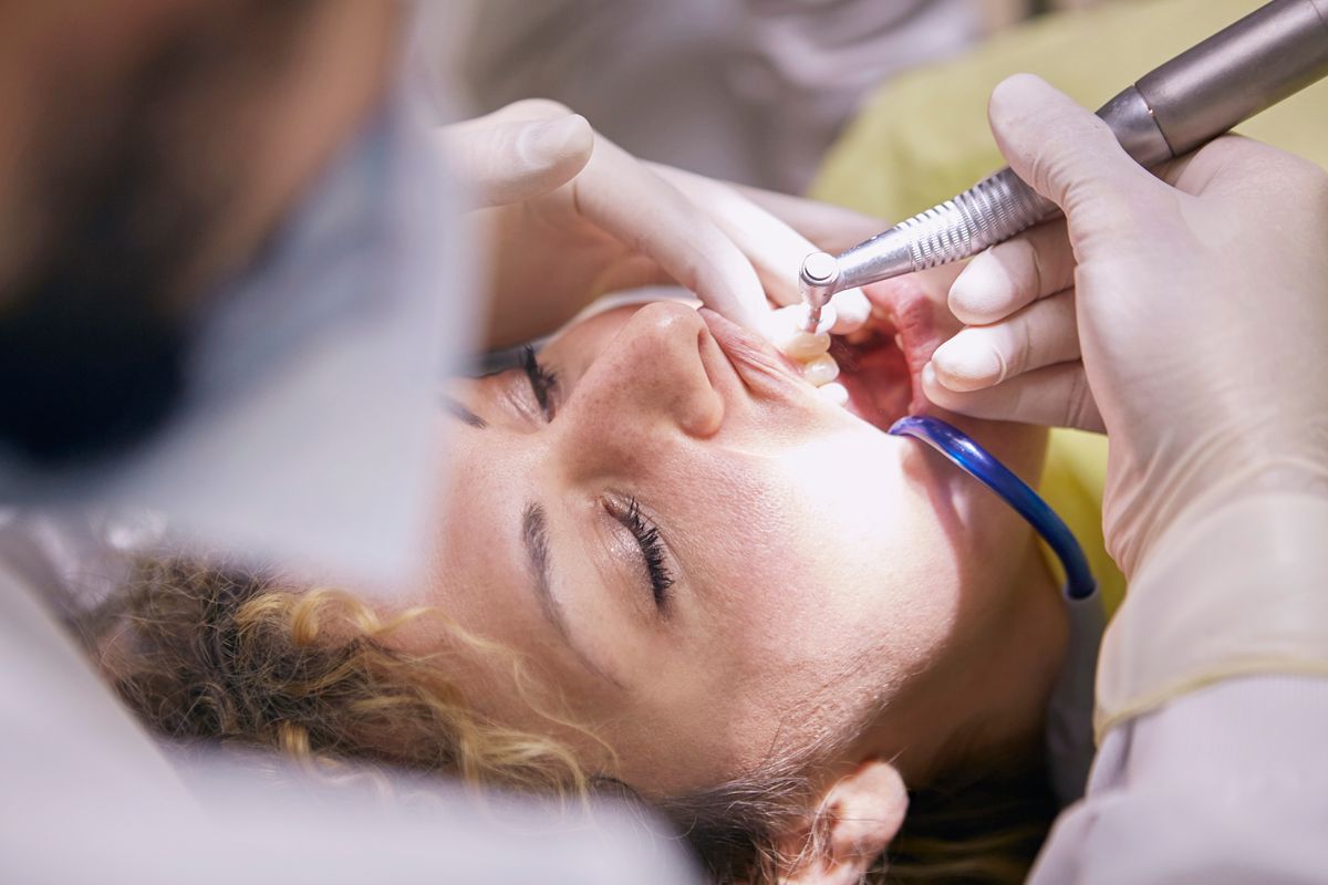
- Phase 5 — Guided Surgery: Print the surgical guide (3D printing or milling), verify fit in the patient's mouth, and perform the osteotomy sequence through the guide sleeves for precise, predictable implant placement.
Key Measurements in CBCT Implant Planning
During the planning phase, several critical measurements determine implant feasibility:
- Available bone height: Distance from the crest to the inferior alveolar canal (mandible) or sinus floor (maxilla). A minimum safety margin of 2 mm from the IAN canal is standard.
- Buccolingual bone width: Measured in cross-sectional views. At least 1 mm of bone should remain buccal and lingual to the implant.
- Bone density: Hounsfield unit (HU) values help predict primary stability and may influence the drilling protocol.
- Interradicular distance: When placing implants between natural teeth, at least 1.5 mm of space from each adjacent root is required.
Flapless vs. Flap Surgery with Guides
CBCT-guided surgery enables flapless (tissue punch) implant placement in appropriate cases. Benefits include less postoperative pain, reduced swelling, faster healing, and preserved blood supply to the crestal bone. However, flapless surgery requires adequate keratinized tissue and confident bone volume assessment — both of which depend on high-quality CBCT data.
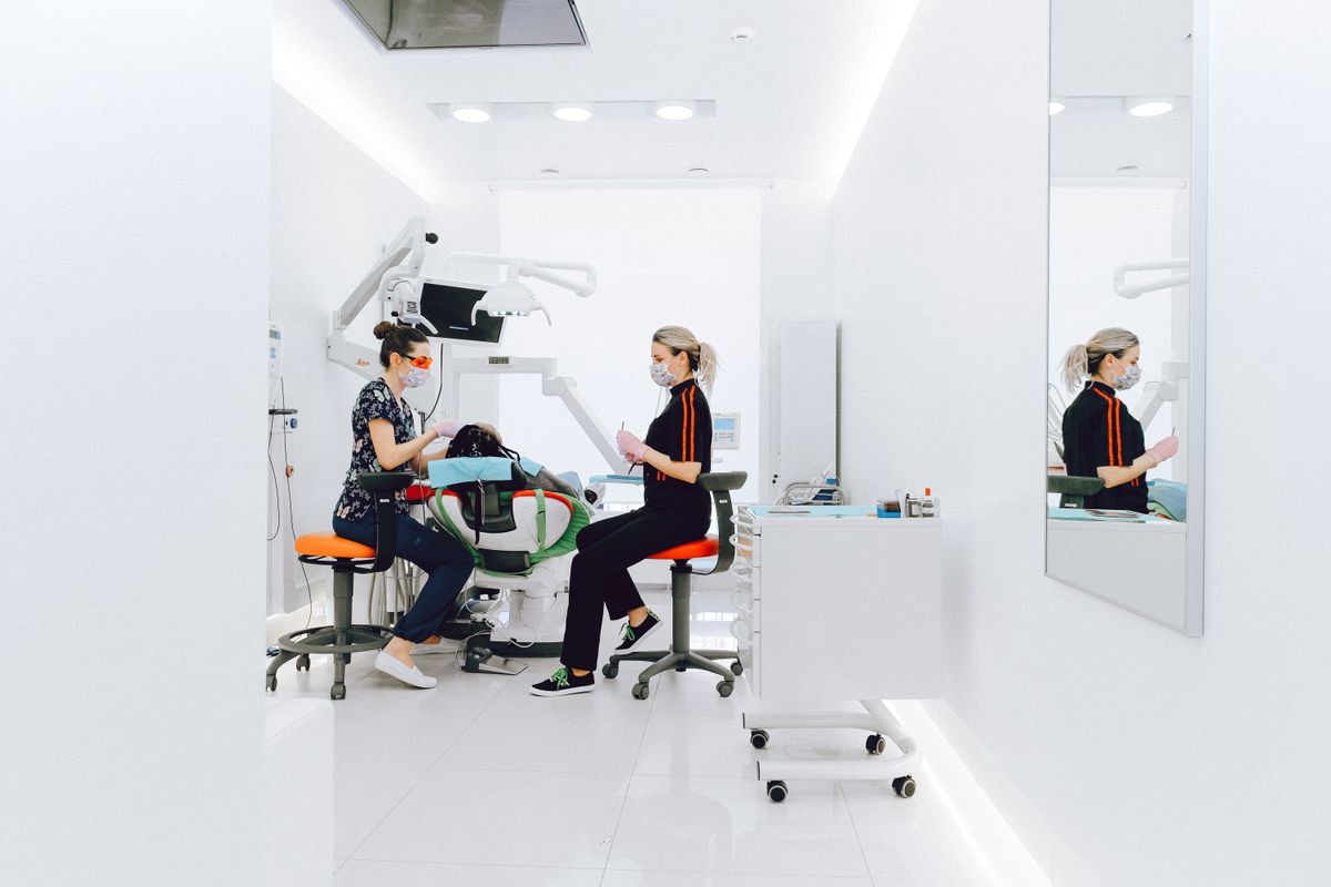
Sharing Plans with the Lab and Referral Network
Digital implant planning is inherently collaborative. The restorative dentist, surgeon, and lab technician all need access to the CBCT data and planned implant positions. With CBCTHub, you can share the CBCT scan via a link, allowing all team members to review the anatomy and discuss the plan — without shipping CDs or requiring everyone to use the same software.
Try CBCTHub for free
Upload, view, and share DICOM scans in the cloud. Nothing to install.
Create free accountRelated articles
Mac vs PC for opening CBCT: what to consider before buying
You need to buy a computer for your clinic or imaging center and are torn between Mac and PC. Real criteria for CBCT, no marketing.

iPad for dental radiology: which model is best in 2026
You want to use iPad to review CBCT scans with patient or referrer. Which model fits best? Honest guide based on real clinical use.
How to migrate from CD to cloud without losing old scans
You have years of CBCT scans on CDs and external drives. Here is a step-by-step plan to migrate to cloud without losing a single one.